PE-PROC-03الإصدار 1.0 · آخر مراجعة أكتوبر 2026 · المراجعة القادمة أكتوبر 2028Version 1.0 · Last reviewed October 2026 · Next review October 2028
يُرجى قراءة هذه النشرة بعناية قبل موعدك وإحضارها معك يوم الفحص. وإذا كان أي أمر غير واضح، تواصل مع وحدة المناظير قبل الموعد لا في يوم الفحص.
ما هو تنظير الأمعاء الدقيقة بالبالونين؟
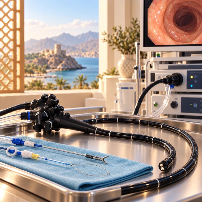
الأمعاء الدقيقة يبلغ طولها عدة أمتار وتقع في التفافات لا يصل إليها المنظار المعتاد. ويستخدم هذا الفحص أنبوباً رفيعاً طويلاً مزوّداً بكاميرا يمرّ داخل أنبوب خارجي، ولكلٍّ منهما بالون لين في طرفه. ويُنفخ البالونان ويُفرغان بالتناوب فتُجمَّع الأمعاء برفق على الأنبوب، مما يتيح للكاميرا الوصول إلى عمق أبعد بكثير من المنظار المعتاد. ويمكن إجراء الفحص عبر الفم أو عبر فتحة الشرج بحسب موضع المشكلة المتوقع، ويستغرق من ٦٠ إلى ٩٠ دقيقة وربما أكثر. ويمكن أخذ العينات، وعند الحاجة إيقاف مواضع النزيف واستئصال السلائل وتوسيع المناطق الضيقة في الجلسة نفسها.
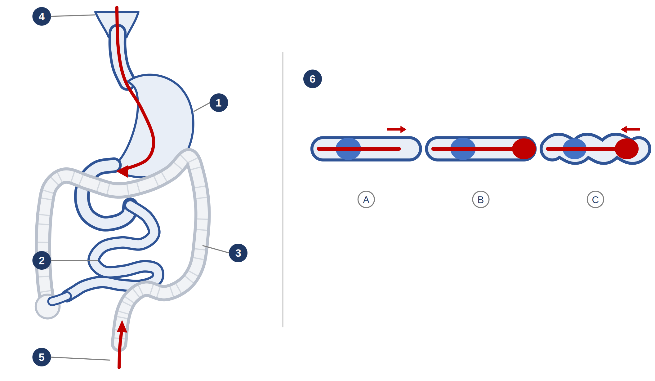
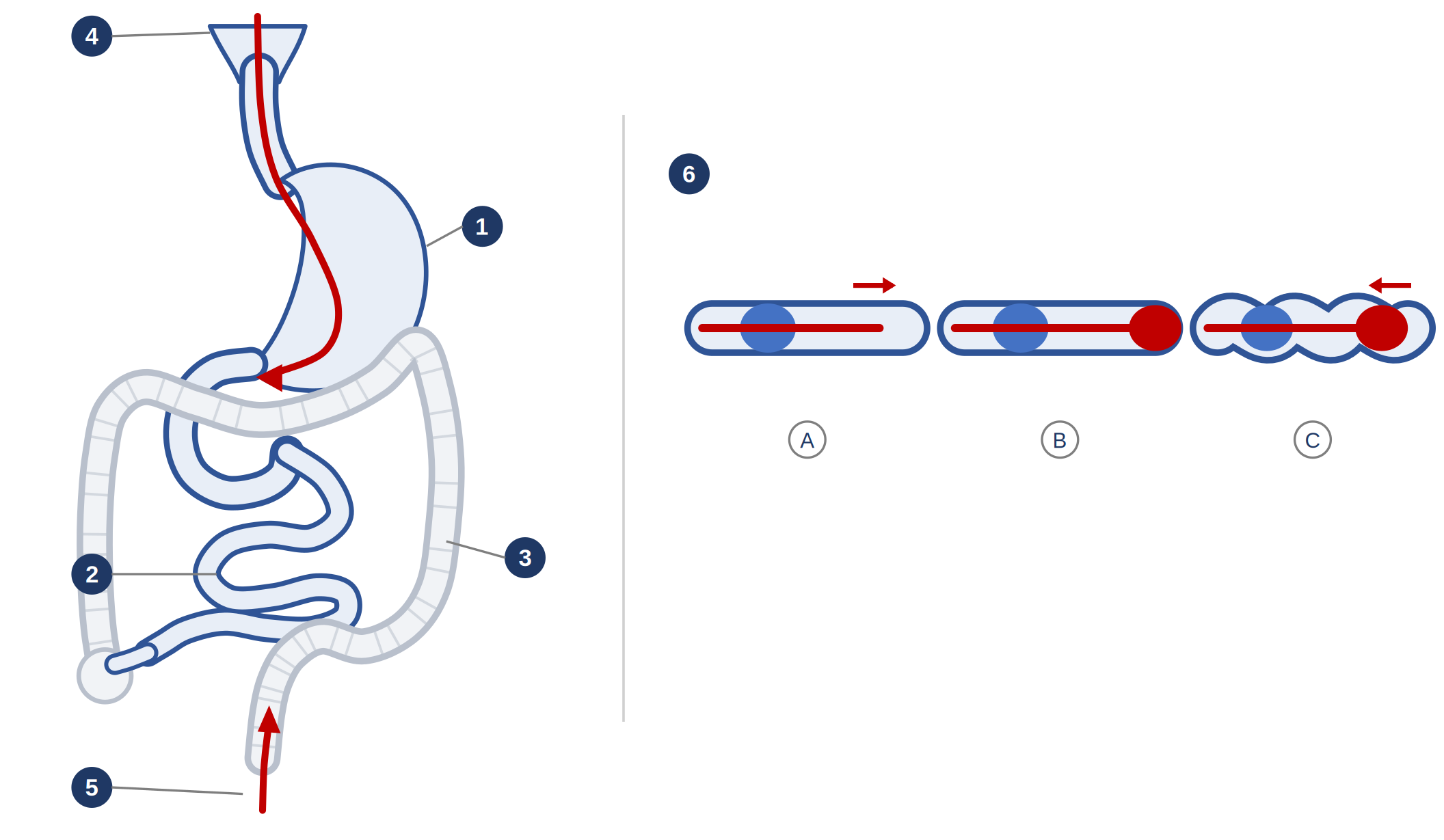
١ المعدة ٢ الأمعاء الدقيقة ٣ القولون ٤ المسار عبر الفم ٥ المسار عبر فتحة الشرج
٦ كيف يعمل البالونان: (أ) يثبّت الأنبوب الخارجي الأمعاء بينما تتقدم الكاميرا · (ب) ينتفخ بالون طرف الكاميرا فيثبّتها · (ج) يُسحب الاثنان للخلف فتتجمّع الأمعاء على الأنبوب
لماذا يُنصح بهذا الفحص؟
أكثر الأسباب شيوعاً هو النزيف أو فقر الدم بنقص الحديد عندما لا يكشف تنظير المعدة وتنظير القولون عن السبب، ويشير فحص الكبسولة أو التصوير إلى الأمعاء الدقيقة. ومن الأسباب الأخرى تقييم أو علاج داء كرون في الأمعاء الدقيقة، واستئصال سلائل الأمعاء الدقيقة، واستخراج كبسولة لم تخرج من الجسم، وفحص القناة الصفراوية لدى المرضى الذين تغيّرت تشريحهم بعمليات جراحية سابقة.
ما فوائده؟
هذا هو الفحص الوحيد الذي يفحص عمق الأمعاء الدقيقة مباشرة ويعالج ما يجده، فيجنّب المريض إجراء عملية جراحية في معظم الحالات. وإذا سبق أن أظهر فحص الكبسولة وجود آفة، فإن التنظير يعثر عليها لدى نحو ثلاثة من كل أربعة مرضى. ويمكن إيقاف النزيف واستئصال السلائل وأخذ العينات لتأكيد التشخيص في جلسة واحدة.
التحضير للفحص
عبر الفم: امتنع عن الطعام الصلب لمدة ثماني ساعات قبل الموعد، وتوقف عن جميع السوائل بما فيها الماء قبل الموعد بأربع إلى ست ساعات حسب ما هو مذكور في خطاب الموعد. وهذه المدد أطول مما هو معتاد في تنظير المعدة، لذا التزم بها بدقة. ولا حاجة إلى تنظيف الأمعاء.
عبر فتحة الشرج: ستُعطى محلول تنظيف الأمعاء كما في تنظير القولون، مع تعليمات مكتوبة منفصلة عن الطعام والتوقيت.
أقراص الحديد: أوقفها قبل الفحص بسبعة أيام.
مميّعات الدم — الوارفارين، الأسبرين، كلوبيدوغريل، تيكاغريلور، براسوغريل، ريفاروكسابان، أبيكسابان، دابيغاتران، إيدوكسابان: تواصل مع الوحدة قبل الموعد بسبعة أيام على الأقل. واستمرارها من عدمه يعتمد على ما إذا كان يُتوقع إجراء علاج أثناء الفحص، لذا يجب أن تصدر التعليمات منّا، ولا توقفها من تلقاء نفسك.
السكري وإبر إنقاص الوزن أو السكري (سيماغلوتايد، ليراغلوتايد، دولاغلوتايد، تيرزيباتايد): أبلغ الوحدة مسبقاً، فهناك تعليمات منفصلة.
رتّب وجود شخص بالغ مسؤول يرافقك إلى المنزل ويبقى معك، وسيُؤجَّل الفحص إذا لم يتوفر مرافق.
التخدير
نظراً لطول مدة الفحص فإنه يُجرى تحت تخدير عميق أو تخدير عام، فتكون نائماً أو مهدّأً بعمق ولن تتذكر شيئاً منه. ويُراقب تنفسك ونبضك وضغط دمك ونسبة الأكسجين طوال الوقت من قِبل فريق مختص. ويغادر معظم المرضى في اليوم نفسه، لكن يُرتَّب المبيت ليلة واحدة إذا أُجري علاج أثناء الفحص أو إذا كانت لديك أمراض أخرى. وخلال أربع وعشرين ساعة بعد الفحص لا تقد السيارة، ولا تسافر بمفردك، ولا تشغّل الآلات، ولا تتناول الكحول، ولا توقّع أوراقاً رسمية، ولا ترعَ أطفالاً صغاراً بمفردك.
ما هي المخاطر؟
يحمل هذا الفحص خطراً أعلى من تنظير المعدة أو القولون المعتاد، لأنه أطول ويصل إلى عمق أبعد. وتحدث المضاعفات الخطيرة في أقل من ١ من كل ١٠٠ فحص بدون علاج، وتصل إلى نحو ٥ من كل ١٠٠ عند إجراء علاج مثل استئصال سليلة أو توسيع منطقة ضيقة.
المضاعفة
مدى تكرارها
انثقاب جدار الأمعاء
نحو ١ من كل ٣٠٠ إلى ١ من كل ١٠٠٠ بدون علاج، وترتفع إلى ١ إلى ٣ من كل ١٠٠ عند استئصال سليلة أو توسيع منطقة ضيقة، وقد تلزم عملية جراحية. وإذا سبق أن أُجريت لك عملية في الأمعاء فالخطر أعلى، وقد سُجّل في المسار عبر فتحة الشرج بنسبة تصل إلى ١ من كل ١٠، وسيُناقش ذلك معك.
التهاب البنكرياس
نحو ١ من كل ٣٠٠، ويحدث في الغالب الأعم بعد المسار عبر الفم. يسبب ألماً وتقيؤاً ويتحسن عادة بإقامة قصيرة في المستشفى. أما ارتفاع تحاليل البنكرياس مؤقتاً دون أعراض فهو أكثر شيوعاً وغير ضار.
النزيف
نحو ١ من كل ٥٠٠ بدون علاج، وأكثر شيوعاً بعد استئصال سليلة. ويتوقف عادة من تلقاء نفسه أو يُعالج أثناء الفحص.
تأثير التخدير على التنفس أو النبض أو ضغط الدم
حتى نحو ١ من كل ٢٠٠. وتتم مراقبتك طوال الفحص وهذه الآثار قابلة للعلاج.
التهاب الحلق والانتفاخ وألم البطن الخفيف
شائعة، نحو ١ من كل ١٠. وهي آثار متوقعة وليست مضاعفة، وتزول خلال ١٢ إلى ٢٤ ساعة.
تعذّر فحص الأمعاء الدقيقة بالكامل
تُفحص الأمعاء الدقيقة بالكامل في نحو الربع إلى النصف من الحالات في الممارسة الأوروبية، حتى عند استخدام المسارين معاً. وكثيراً ما يُرتَّب فحص ثانٍ من الجهة الأخرى، وهو أمر مخطط له وليس مضاعفة.
الوفاة المرتبطة مباشرة بالإجراء
نادرة جداً. لم تُسجَّل أي وفاة في سلسلة دولية ضمت ٢٣٦٢ إجراءً، وإن كانت وفيات متفرقة قد سُجّلت في سجلات أكبر، غالباً بعد انثقاب أو التهاب بنكرياس شديد.
هل توجد بدائل؟
يلتقط فحص الكبسولة صوراً للأمعاء الدقيقة لكنه لا يسمح بأخذ عينات أو بالعلاج، وقد تعلق الكبسولة أحياناً. أما التصوير المقطعي أو بالرنين للأمعاء فيُظهر جدار الأمعاء وما حولها لكن لا يُظهر البطانة بالتفصيل ولا يتيح العلاج. ويبقى إدخال المنظار أثناء عملية جراحية خياراً أخيراً وهو أكثر بضعاً بكثير. كما أن عدم إجراء الفحص خيار مطروح، وينبغي أن تناقش مع طبيبك ما قد يفوتك حينها.
بعد الفحص
ستبقى للإفاقة في الجناح أو غرفة الإفاقة بضع ساعات. والانتفاخ والغازات وألم البطن الخفيف أمور متوقعة تخف بالمشي. ويُسمح عادة بالأكل والشرب بعد استيقاظك تماماً ما لم يُنصح بغير ذلك. وتُشرح لك النتائج قبل مغادرتك ويُسلَّم تقرير مكتوب، وبسبب التخدير اطلب إعادة الشرح أو إعطاءه للشخص المرافق. أما نتائج العينات فتستغرق من أسبوعين إلى أربعة أسابيع وتُناقش في العيادة.
متى تطلب المساعدة العاجلة
توجّه إلى أقرب قسم للطوارئ إذا ظهر عليك خلال يومين من الفحص أي مما يلي:
ألم بطني شديد، خاصة إذا ازداد بالحركة أو السعال، أو انتفاخ وتصلّب في البطن
ألم في أعلى الكتف
تقيؤ مستمر أو تقيؤ دم
نزيف غزير أو متكرر من فتحة الشرج، أو تجلطات، أو براز أسود
ارتفاع الحرارة إلى ٣٨ درجة مئوية أو أكثر، أو صعوبة في التنفس
خذ هذه النشرة معك وأخبر الفريق الطبي بأنك خضعت لتنظير الأمعاء الدقيقة بالبالونين.
للتواصل
للمواعيد والاستفسارات، تواصل مع وحدة المناظير عبر الأرقام التي زوّدتك بها. وإذا كانت حالتك شديدة أو كان الوقت خارج أوقات العمل فتوجّه إلى قسم الطوارئ بدلاً من انتظار الرد.
أُعدّت استناداً إلى إرشادات الجمعية الأوروبية لتنظير الجهاز الهضمي الخاصة بتنظير الأمعاء الدقيقة ومعايير أدائها، وإرشادات مميّعات الدم البريطانية والأوروبية، وبيانات السلامة المنشورة من دراسات متعددة المراكز. وهي تدعم مناقشتك مع طبيبك وإجراءات الموافقة ولا تحل محلها. PE-PROC-03 · الإصدار ١٫٠ · أكتوبر ٢٠٢٦ · المراجعة: أكتوبر ٢٠٢٨
هذه النشرة معلومات عامة ولا تغني عن مشورة طبيبك المعالج.
زاد صحي، PE-PROC-03
Please read this sheet carefully before your appointment and bring it with you on the day. If anything is unclear, contact the Endoscopy Unit before your procedure rather than on the day.
What is a double balloon enteroscopy?
The small bowel is several metres long and lies in loops that an ordinary endoscope cannot reach. A double balloon enteroscopy uses a long, thin camera tube inside an outer tube, each with a soft balloon at its tip. The balloons are inflated and deflated in turn so that the bowel is gently gathered over the tube, allowing the camera to travel far deeper than a standard endoscopy. The examination can be done through the mouth or through the back passage, depending on where the problem is thought to be, and takes 60 to 90 minutes — sometimes longer. Biopsies can be taken and, where needed, bleeding points treated, polyps removed and narrowed segments stretched during the same procedure.
1 Stomach 2 Small bowel 3 Colon 4 Route through the mouth 5 Route through the back passage
6 How the balloons work: A the outer tube grips the bowel while the camera advances · B the camera tip balloon grips · C both are drawn back, gathering the bowel over the tube
Why has it been recommended for me?
The commonest reason is bleeding or iron deficiency anaemia when a gastroscopy and colonoscopy have not found the cause and a capsule endoscopy or scan has pointed to the small bowel. Other reasons include assessment or treatment of Crohn’s disease of the small bowel, removal of small bowel polyps, retrieval of a capsule that has not passed, and examination of the bile duct in patients whose anatomy has been altered by previous surgery.
What are the benefits?
This is the only test that both examines the deep small bowel directly and treats what it finds, avoiding an operation in most cases. When a capsule endoscopy has already shown a lesion, an enteroscopy finds it in about three out of four patients. Bleeding points can be sealed, polyps removed and biopsies taken to confirm a diagnosis, all in one procedure.
Preparing for the test
Through the mouth: do not eat solid food for 8 hours before your appointment, and stop all fluids including water 4 to 6 hours before, as stated in your appointment letter. These times are longer than for a routine gastroscopy, so please follow them exactly. No bowel preparation is needed.
Through the back passage: you will be given bowel preparation to take, exactly as for a colonoscopy, along with separate written instructions on diet and timing.
Iron tablets: stop 7 days before the test.
Blood thinners — warfarin, aspirin, clopidogrel, ticagrelor, prasugrel, rivaroxaban, apixaban, dabigatran, edoxaban: contact the unit at least 7 days before. Whether they are continued depends on whether treatment is planned during the procedure, so the instruction must come from us. Do not stop them on your own.
Diabetes, and weight-loss or diabetes injections (semaglutide, liraglutide, dulaglutide, tirzepatide): tell the unit in advance, as separate instructions are needed.
Arrange for a responsible adult to take you home and stay with you. The procedure will be postponed if no escort is available.
Sedation and anaesthesia
Because the procedure is long, it is done under deep sedation or a general anaesthetic. You will be asleep or heavily sedated and will not remember it. Your breathing, heart rate, blood pressure and oxygen level are monitored throughout by a dedicated team. Most patients go home the same day, but an overnight stay is arranged if treatment was carried out during the procedure or if you have other medical conditions. For 24 hours afterwards you must not drive, travel alone, operate machinery, drink alcohol, sign legal documents, or care for young children on your own.
What are the risks?
Enteroscopy carries a higher risk than a standard gastroscopy or colonoscopy, because the examination is longer and reaches much further. Serious complications occur in fewer than 1 in 100 examinations without treatment, and in up to about 5 in 100 when treatment such as polyp removal or stretching a narrowing is carried out.
Complication
How often it happens
Perforation (a tear in the bowel wall)
About 1 in 300 to 1 in 1,000 without treatment, rising to 1 to 3 in 100 when a polyp is removed or a narrowing is stretched. Surgery may be needed. If you have had previous bowel surgery the risk is higher — for the route through the back passage it has been reported as high as 1 in 10, and this will be discussed with you.
Inflammation of the pancreas (pancreatitis)
About 1 in 300, and almost always after the route through the mouth. It causes pain and vomiting and usually settles with a short hospital stay. A temporary rise in pancreas blood tests without any symptoms is much more common and harmless.
Bleeding
About 1 in 500 without treatment, and more common after a polyp is removed. It usually stops on its own or is treated during the procedure.
Effects of sedation or anaesthesia on breathing, heart rate or blood pressure
Up to about 1 in 200. You are monitored throughout and these effects can be treated.
Sore throat, bloating and abdominal discomfort
Common — about 1 in 10. These are expected effects rather than complications and settle within 12 to 24 hours.
The whole small bowel cannot be examined
In European practice the whole small bowel is seen in roughly a quarter to a half of examinations, even when both routes are used. A second procedure from the other end is often arranged, and this is planned rather than a complication.
Death directly related to the procedure
Very rare. No deaths occurred in an international series of 2,362 procedures, although occasional deaths have been reported in larger registries, usually after a perforation or severe pancreatitis.
Are there alternatives?
Capsule endoscopy takes pictures of the small bowel but cannot take biopsies or treat anything, and the capsule occasionally becomes stuck. CT or MR enterography shows the wall and structures around the bowel but not the lining in detail, and again allows no treatment. An operation with the endoscope passed during surgery is the remaining option and is far more invasive. Choosing not to have the test is also an option; discuss with your doctor what might be missed.
After the procedure
You will recover in the ward or recovery area for a few hours. Bloating, wind and mild abdominal discomfort are expected and are relieved by walking. Eating and drinking are usually allowed once you are fully awake, unless you are told otherwise. The findings are explained before you leave and a written report is given; because of the sedation, ask for the information to be repeated or given to the person accompanying you. Biopsy results take 2 to 4 weeks and are discussed in clinic.
When to get urgent help
Go to the nearest emergency department if, within two days of the test, you develop:
severe abdominal pain, particularly pain made worse by moving or coughing, or a swollen, hard abdomen
pain at the tip of the shoulder
persistent vomiting, or vomiting blood
heavy or repeated rectal bleeding, clots, or black tarry stools
fever of 38 °C or above, or difficulty breathing
Take this sheet with you and tell the staff that you have had a double balloon enteroscopy.
Contact
For appointments and questions, contact the endoscopy unit using the numbers it has given you. If you are seriously unwell, or it is outside working hours, go to the Emergency Department rather than waiting for a reply.
Based on ESGE small bowel endoscopy guidelines, BSG/ESGE antithrombotic guidance and published multicentre enteroscopy safety data. Supports but does not replace the discussion with your doctor and the consent process. PE-PROC-03 · Version 1.0 · October 2026 · Review due: October 2028
This leaflet is general information and does not replace advice from your own doctor.