PE-PROC-05الإصدار 1.0 · آخر مراجعة أكتوبر 2026 · المراجعة القادمة أكتوبر 2028Version 1.0 · Last reviewed October 2026 · Next review October 2028
يُرجى قراءة هذه النشرة بعناية قبل موعدك وإحضارها معك يوم الفحص. وإذا كان أي أمر غير واضح، تواصل مع وحدة المناظير قبل الموعد لا في يوم الفحص.
ما هو تنظير الكبسولة؟
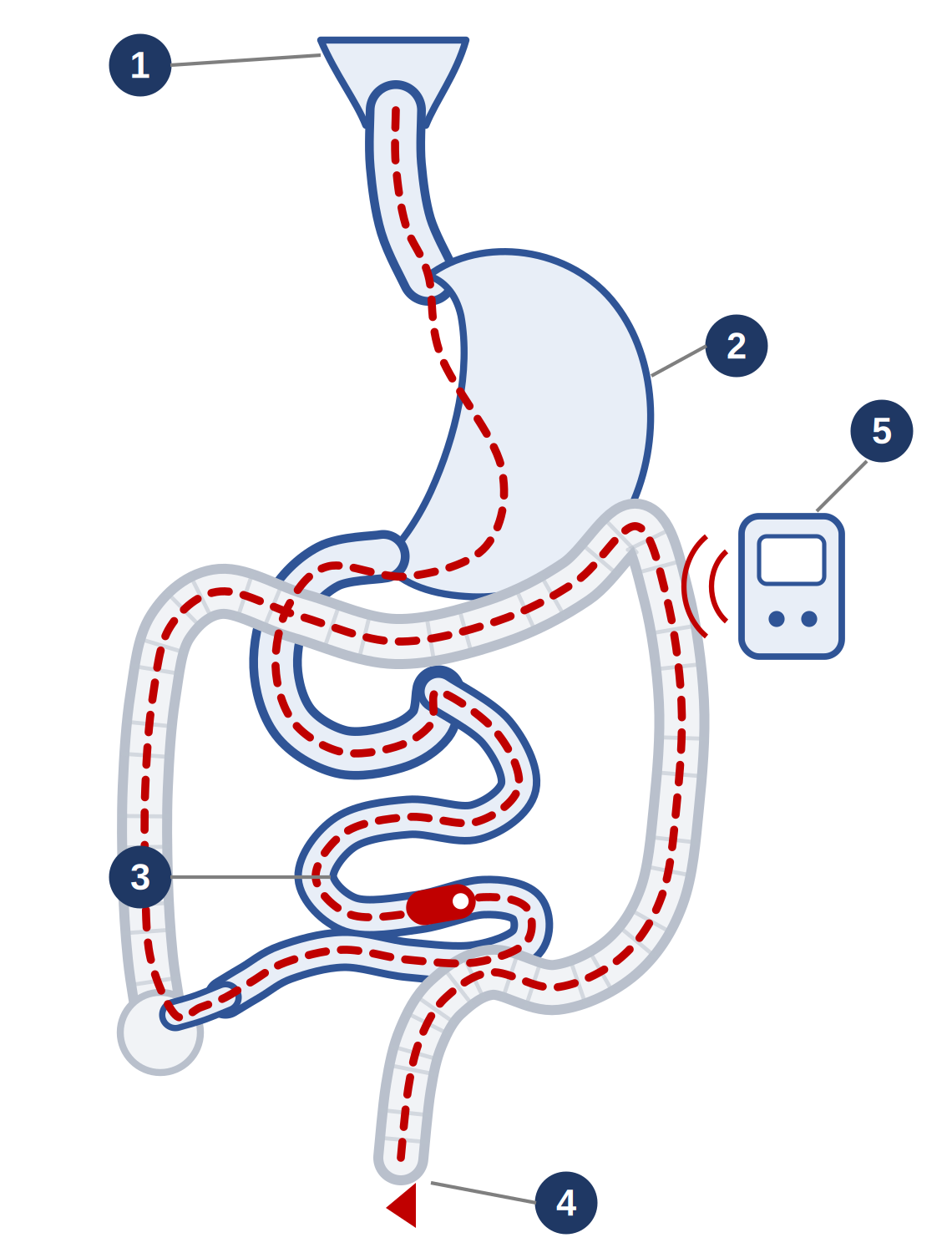
يستخدم هذا الفحص كاميرا بحجم حبة دواء كبيرة تبتلعها مع الماء. وبينما تسير في الجهاز الهضمي تلتقط عشرات الآلاف من الصور للأمعاء الدقيقة وترسلها إلى جهاز تسجيل يُحمل على حزام حول الخصر. ولا يوجد أنبوب ولا تخدير. ولا تُستخرج الكبسولة، بل تخرج طبيعياً مع البراز خلال يوم أو يومين عادة، ولا يُعاد استخدامها. وهو الوسيلة المريحة الوحيدة لرؤية الأمعاء الدقيقة بكاملها، وهي تمتد عدة أمتار ولا يصل إليها تنظير المعدة ولا تنظير القولون.
١ ابتلاع الكبسولة ٢ المعدة ٣ الأمعاء الدقيقة — وهي موضع الفحص ٤ القولون حيث تخرج الكبسولة طبيعياً ٥ جهاز التسجيل على الحزام
لماذا يُنصح بهذا الفحص؟
أكثر الأسباب شيوعاً فقر الدم بنقص الحديد أو النزيف عندما لا يكشف تنظير المعدة وتنظير القولون عن السبب، والاشتباه بداء كرون في الأمعاء الدقيقة أو متابعته. ويُستخدم أيضاً في مرض السيلياك الذي لا يستجيب للحمية الخالية من الغلوتين، وفي أمراض السلائل الوراثية.
ما الذي يستطيعه هذا الفحص وما لا يستطيعه
تلتقط الكبسولة الصور فقط؛ فهي لا تأخذ عينات ولا توقف نزيفاً ولا تستأصل شيئاً، ولا يمكن توجيهها أو إيقافها. وفي المرضى الذين يُفحصون لنزيف الأمعاء الدقيقة تكشف عن سبب محتمل لدى نحو ٦ من كل ١٠. وفي فقر الدم بنقص الحديد تُكتشف آفة لدى نحو النصف، وآفة تفسّر فقر الدم لدى نحو ٤ من كل ١٠. وإذا وُجد ما يحتاج إلى عينة أو علاج فيُرتَّب عادة تنظير الأمعاء بالبالونين بعده. أما إذا اكتمل الفحص وكان طبيعياً فلا يحتاج معظم المرضى إلى فحص إضافي، وإن كان نحو ١ من كل ٥ ممن فُحصوا بسبب النزيف يعاودهم النزيف ويحتاجون إلى إعادة تقييم.
قبل الفحص: هل تلزم كبسولة اختبار المرور؟
إذا كان لديك داء كرون معروف، أو أعراض توحي بوجود تضيّق، أو سبق أن أُجريت لك عملية في الأمعاء، أو تلقيت علاجاً إشعاعياً للبطن، أو تتناول مسكّنات مضادة للالتهاب بانتظام، فقد تُعطى أولاً كبسولة اختبار المرور. وهي كبسولة صمّاء بالحجم نفسه مصنوعة من مادة تتفكك من تلقاء نفسها بعد نحو ثلاثين ساعة من ابتلاعها. فإذا خرجت سليمة أمكن إعطاء الكبسولة الحقيقية بأمان، وهذا يقلل احتمال احتباسها. ونادراً ما تسبب كبسولة الاختبار نفسها مغصاً أو أعراض انسداد؛ فأخبر وحدة المناظير إن حدث ذلك.
التحضير للفحص
اتبع تعليمات الطعام وتنظيف الأمعاء التي يعطيك إياها فريق المناظير. فنظافة الأمعاء الدقيقة هي ما يجعل الصور صالحة للقراءة.
امتنع عن الأكل والشرب ابتداءً من منتصف الليل في الليلة السابقة، ما لم يُنصح بغير ذلك.
أقراص الحديد: أوقفها قبل الفحص بسبعة أيام، لأن الحديد يترك بقايا داكنة في الأمعاء تحجب الرؤية.
تُستمر مميّعات الدم عادة لأنه لا تُؤخذ عينات في هذا الفحص. ولا توقف أي مميّع للدم إلا إذا طلبنا منك ذلك.
أبلغ الوحدة مسبقاً إذا كان لديك جهاز تنظيم لضربات القلب أو مزيل رجفان مزروع، أو صعوبة في بلع الأقراص، أو سبق أن أُجريت لك عملية في الأمعاء أو علاج إشعاعي للبطن، أو كنتِ حاملاً أو يُحتمل ذلك، إذ لا يُجرى الفحص عادة أثناء الحمل. وإذا كنت مصاباً بالسكري فأبلغ وحدة المناظير كذلك ليُعدَّل الإنسولين أو الأقراص حول فترة الصيام.
ارتدِ ملابس واسعة من قطعتين، إذ يُحمل جهاز التسجيل على حزام نحو ٨ إلى ١٢ ساعة.
يوم الفحص
يُركَّب جهاز التسجيل ثم تبتلع الكبسولة مع الماء، وبعدها تغادر الوحدة وتعود في وقت لاحق من اليوم لنزع الجهاز، ولا تحتاج إلى البقاء في المستشفى. وامتنع عن الأكل والشرب لمدة ساعتين بعد ابتلاع الكبسولة، ثم يمكنك تناول السوائل الصافية، ووجبة خفيفة بعد أربع ساعات. وتجنّب المجهود البدني الشديد والانحناء لفترات طويلة أثناء ارتداء الجهاز، ولا تفصله. وإذا كنت تجد صعوبة في بلع الأقراص فأخبر وحدة المناظير، إذ يمكن وضع الكبسولة مباشرة في المعدة بمنظار قصير.
ما هي المخاطر؟
تنظير الكبسولة أقل فحوصات الأمعاء الدقيقة بضعاً؛ فلا تخدير فيه ولا أنبوب ولا خطر تمزق من أداة. والخطر الرئيسي هو عدم مرور الكبسولة عبر منطقة ضيقة.
الخطر
مدى تكراره
عدم خروج الكبسولة (احتباس الكبسولة)
نحو ١ إلى ٢ من كل ١٠٠ فحص إجمالاً؛ ونحو ٢ من كل ١٠٠ عند الاشتباه بداء كرون، ونحو ٥ من كل ١٠٠ — وتصل إلى ٨ من كل ١٠٠ في تقارير أقدم — لدى المصابين بداء كرون. وتقلل كبسولة اختبار المرور هذه النسبة.
ماذا يحدث إذا لم تخرج
لا تظهر أعراض على معظم المرضى، وتخرج الكبسولة من تلقاء نفسها أو بعد علاج التضيّق الذي أعاقها. وإن لم تخرج فيمكن استخراجها عادة بتنظير الأمعاء بالبالونين. ونحو ثلث حالات الاحتباس تنتهي بعملية جراحية، أي نحو ٥ إلى ٧ من كل ١٠٠٠ فحص، وتُجرى غالباً بسبب التضيّق نفسه لا من أجل الكبسولة.
أعراض الانسداد
غير شائعة. وإذا احتبست الكبسولة فقد تسبب ألماً في البطن وتقيؤاً وانتفاخاً، وهي حالة تستدعي تقييماً عاجلاً.
دخول الكبسولة إلى مجرى الهواء بدلاً من المريء
نادر، نحو ١ من كل ١٠٠٠، ويحدث في الغالب لدى كبار السن أو من لديهم صعوبة في البلع. فأخبر وحدة المناظير مسبقاً إن كنت تجد صعوبة في بلع الأقراص، إذ يمكن حينها وضع الكبسولة بمنظار قصير.
عدم اكتمال الفحص
في نحو ١ من كل ١٠ فحوص للمرضى الخارجيين تنفد البطارية قبل وصول الكبسولة إلى الأمعاء الغليظة، فلا يُرى جزء من الأمعاء الدقيقة. وهو أكثر حدوثاً لدى المنوّمين وكبار السن، وقد يستلزم إعادة الفحص.
رداءة الرؤية
إذا لم تكن الأمعاء نظيفة بما يكفي فقد تفوت آفات صغيرة ويلزم إعادة الفحص، ولهذا يُعدّ التحضير مهماً.
هل توجد بدائل؟
يُظهر التصوير المقطعي أو بالرنين المغناطيسي للأمعاء جدار الأمعاء والأنسجة المحيطة به وهو أفضل في كشف التضيّقات، لكنه لا يُظهر البطانة بالتفصيل. أما تنظير الأمعاء بالبالونين فيفحص البطانة ويسمح بأخذ العينات وبالعلاج، لكنه فحص باضع يستغرق من ٦٠ إلى ٩٠ دقيقة ويحتاج إلى تخدير عميق. وهذه الفحوص يكمّل بعضها بعضاً عملياً، وتُجرى الكبسولة أولاً في العادة.
بعد الفحص
عُد في الوقت المحدد لنزع جهاز التسجيل، ثم تناول الطعام والشراب كالمعتاد. وتابع البراز حتى ترى الكبسولة؛ فمعظمها يخرج خلال يوم أو يومين، وقد يستغرق الأمر أسبوعين دون أن يكون ذلك غير طبيعي بالضرورة. ولا تُجرِ تصويراً بالرنين المغناطيسي قبل أن تتأكد من خروجها، وإن لم تكن متأكداً فتواصل مع وحدة المناظير ويُجرى تصوير بالأشعة للتأكد. وإذا لم ترها خلال أسبوعين فتواصل مع الوحدة. ويحتوي التسجيل على آلاف الصور ويستغرق وقتاً لمراجعته، وتُناقش النتائج معك في العيادة.
متى تطلب المساعدة العاجلة
توجّه إلى أقرب قسم للطوارئ إذا ظهر عليك في أي وقت بعد ابتلاع الكبسولة أي مما يلي:
ألم في البطن شديد أو متزايد، أو انتفاخ في البطن
تقيؤ متكرر أو عدم القدرة على الاحتفاظ بالسوائل
عدم القدرة على التبرز أو إخراج الغازات
سعال مفاجئ أو اختناق أو ضيق في التنفس بعد ابتلاع الكبسولة
أخبر الفريق الطبي بأنك ابتلعت كبسولة تصوير وأطلعهم على هذه النشرة.
للتواصل
للمواعيد والاستفسارات، تواصل مع وحدة المناظير عبر الأرقام التي زوّدتك بها. وإذا كانت حالتك شديدة أو كان الوقت خارج أوقات العمل فتوجّه إلى قسم الطوارئ بدلاً من انتظار الرد.
أُعدّت استناداً إلى إرشادات الجمعية الأوروبية لتنظير الجهاز الهضمي الخاصة بالأمعاء الدقيقة ومعايير أدائها، وتقييم التقنية الصادر عن الجمعية الأمريكية لتنظير الجهاز الهضمي، والبيانات المنشورة حول الاحتباس ونسب الكشف. وهي تدعم مناقشتك مع طبيبك وإجراءات الموافقة ولا تحل محلها. PE-PROC-05 · الإصدار ١٫٠ · أكتوبر ٢٠٢٦ · المراجعة: أكتوبر ٢٠٢٨
هذه النشرة معلومات عامة ولا تغني عن مشورة طبيبك المعالج.
زاد صحي، PE-PROC-05
Please read this sheet carefully before your appointment and bring it with you on the day. If anything is unclear, contact the Endoscopy Unit before your procedure rather than on the day.
What is a capsule endoscopy?
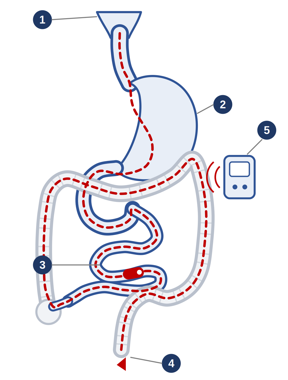
A capsule endoscopy uses a camera the size of a large tablet, which you swallow with water. As it travels through the digestive system it takes tens of thousands of pictures of the small bowel and sends them to a recorder worn on a belt around your waist. There is no tube and no sedation. The capsule is not retrieved: it passes out naturally with a bowel motion, usually within a day or two, and is not reused. It is the only comfortable way to see the whole small bowel, which is several metres long and out of reach of a gastroscopy or colonoscopy.
1 Capsule swallowed 2 Stomach 3 Small bowel — the part this test examines 4 Colon, where the capsule passes out naturally 5 Recorder worn on a belt
Why has it been recommended for me?
The commonest reasons are iron deficiency anaemia or bleeding when a gastroscopy and colonoscopy have not found the cause, and suspected or known Crohn’s disease of the small bowel. It is also used in coeliac disease that is not responding to a gluten-free diet, and in inherited polyp conditions.
What it can and cannot do
The capsule takes pictures only. It cannot take biopsies, stop bleeding or remove anything, and it cannot be steered or held still. In patients investigated for small bowel bleeding it finds a likely cause in roughly 6 out of 10. In iron deficiency anaemia a lesion is found in about half, and one that explains the anaemia in about 4 out of 10. If something is found that needs a biopsy or treatment, a balloon enteroscopy is usually arranged afterwards. If the examination is complete and normal, most patients need no further small bowel test, although about 1 in 5 of those investigated for bleeding will bleed again and need review.
Before the test: is a patency capsule needed first?
If you have established Crohn’s disease, symptoms suggesting a narrowing, previous bowel surgery, previous radiotherapy to the abdomen, or take anti-inflammatory painkillers regularly, you may first be given a patency capsule. This is a dummy capsule of the same size, made of material that breaks down by itself about 30 hours after you swallow it. If it passes whole, a real capsule can be given safely, and this lowers the chance of the capsule becoming stuck. Rarely the patency capsule itself causes cramping or obstructive symptoms — tell the endoscopy unit if that happens.
Preparing for the test
Follow the diet and bowel preparation instructions given to you by the endoscopy staff. A clean small bowel is what makes the pictures readable.
Do not eat or drink from midnight the night before, unless told otherwise.
Iron tablets: stop 7 days before, as iron leaves a dark residue in the bowel that obscures the view.
Blood thinners are usually continued, as no biopsies are taken. Do not stop any blood thinner unless we tell you to.
Tell the unit in advance if you have a pacemaker or implanted defibrillator, any difficulty swallowing tablets, previous bowel surgery or radiotherapy, or if you are or might be pregnant — capsule endoscopy is not normally performed in pregnancy. If you have diabetes, tell the endoscopy unit as well, so your insulin or tablets can be adjusted around the fasting.
Wear loose, two-piece clothing. The recorder is worn on a belt for about 8 to 12 hours.
On the day
The recorder is fitted and you swallow the capsule with water. You then leave the department and return later in the day to have the recorder removed — you do not stay in hospital. Do not eat or drink for 2 hours after swallowing the capsule. You may then take clear fluids, and a light meal after 4 hours. Avoid strenuous exercise and bending for long periods while the recorder is on, and do not disconnect it. If you have difficulty swallowing tablets, tell the endoscopy unit: the capsule can be placed directly into the stomach with a short endoscopy.
What are the risks?
Capsule endoscopy is the least invasive of the small bowel tests. There is no sedation, no tube and no risk of a tear from an instrument. The main risk is that the capsule does not pass through a narrowed segment.
Risk
How often it happens
The capsule does not pass (capsule retention)
About 1 to 2 in 100 examinations overall; about 2 in 100 if Crohn’s disease is suspected, and about 5 in 100 — up to 8 in 100 in older reports — if you already have Crohn’s disease. A patency capsule beforehand lowers this.
What happens if it does not pass
Most patients have no symptoms, and the capsule passes on its own or after treatment of the narrowing that held it up. If it does not, it can usually be retrieved at a balloon enteroscopy. About a third of retained capsules end in an operation — roughly 5 to 7 in every 1,000 examinations — and this is often done for the narrowing itself rather than for the capsule.
Blockage symptoms
Uncommon. If the capsule sticks it can cause abdominal pain, vomiting and a swollen abdomen, which need urgent assessment.
The capsule going into the airway instead of the food pipe
Rare — about 1 in 1,000, and almost always in older patients or those with difficulty swallowing. Tell the endoscopy unit beforehand if you have any trouble swallowing tablets, as the capsule can then be placed with a short endoscopy instead.
Incomplete examination
In about 1 in 10 outpatient examinations the battery runs out before the capsule reaches the large bowel, so part of the small bowel is not seen. It is commoner in inpatients and in older patients. The test may need repeating.
Poor views
If the bowel is not clean enough, small lesions can be missed and the test may have to be repeated. This is why the preparation matters.
Are there alternatives?
CT or MR enterography shows the bowel wall and the tissues around it and is better at finding narrowings, but it does not show the lining in detail. Balloon enteroscopy examines the lining and can take biopsies and treat what it finds, but it is invasive, takes 60 to 90 minutes and needs deep sedation. In practice these tests complement each other, and the capsule is usually done first.
After the test
Return as instructed to have the recorder removed, then eat and drink normally. Check your bowel motions until you see the capsule. Most pass within a day or two, but up to two weeks is not in itself abnormal. Until you are sure it has passed, do not have an MRI scan; if you are unsure, contact the endoscopy unit and a plain X-ray will confirm. If you have not seen it within two weeks, contact the unit. The recording contains many thousands of images and takes time to review; the results are discussed with you in clinic.
When to get urgent help
Go to the nearest emergency department if, at any time after swallowing the capsule, you develop:
abdominal pain that is severe or getting worse, or a swollen abdomen
repeated vomiting, or being unable to keep fluids down
being unable to open your bowels or pass wind
sudden coughing, choking or breathlessness after swallowing the capsule
Tell the staff that you have swallowed a capsule camera and show them this sheet.
Contact
For appointments and questions, contact the endoscopy unit using the numbers it has given you. If you are seriously unwell, or it is outside working hours, go to the Emergency Department rather than waiting for a reply.
Based on ESGE small bowel endoscopy guidance and performance measures, ASGE technology assessment, and published retention and yield data. Supports but does not replace the discussion with your doctor and the consent process. PE-PROC-05 · v1.0 · October 2026 · Review: October 2028
This leaflet is general information and does not replace advice from your own doctor.