PE-PROC-08الإصدار 1.0 · آخر مراجعة أكتوبر 2026 · المراجعة القادمة أكتوبر 2028Version 1.0 · Last reviewed October 2026 · Next review October 2028
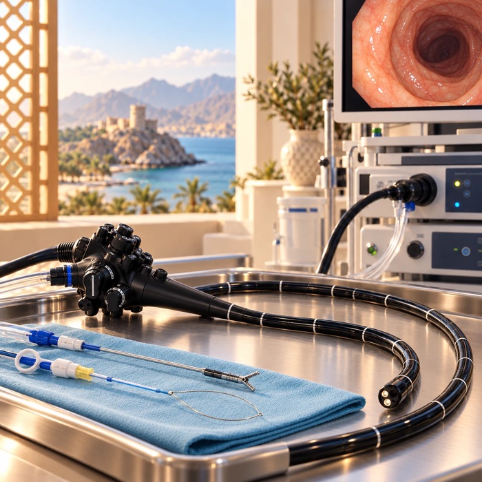
فحص بالكاميرا مزوّد بمسبار للموجات فوق الصوتية في طرفه. ولأن المسبار يكون داخل الجهاز الهضمي ملاصقاً للعضو المراد فحصه، فإنه يعطي صورة أدق بكثير من التصوير من خارج الجسم.
ما هو الإجراء ولماذا تحتاجه
يُدخل أنبوب رفيع مرن عبر الفم كما في تنظير المعدة المعتاد، غير أن هذا يحمل مسباراً صغيراً للموجات فوق الصوتية في نهايته. ومن داخل المعدة وبداية الأمعاء الدقيقة يستطيع المسبار رؤية البنكرياس والقناة الصفراوية وجدار المريء والمعدة والغدد والتراكيب المجاورة لها مباشرة.
ويُستخدم لفحص كتلة أو كيس في البنكرياس، ولتحديد مدى انتشار الورم قبل التخطيط للعلاج، وللبحث عن حصوات صغيرة في القناة الصفراوية قد تفوتها الأشعة، ولفحص كتلة نامية في جدار المريء أو المعدة.
ويمكن تمرير إبرة رفيعة عبر المنظار وجدار الأمعاء لأخذ عينات من كتلة أو غدة، أو لسحب سائل من كيس. ويسمى ذلك السحب أو الخزعة بالإبرة الدقيقة، ويُجرى بتوجيه الموجات فوق الصوتية فتُراقَب الإبرة طوال مسارها. وإذا سُحب سائل من كيس فيُعطى مضاد حيوي أيضاً في العادة. وليس كل فحص يتضمن إبرة، وسنخبرك مسبقاً إن كان ذلك مخططاً له.
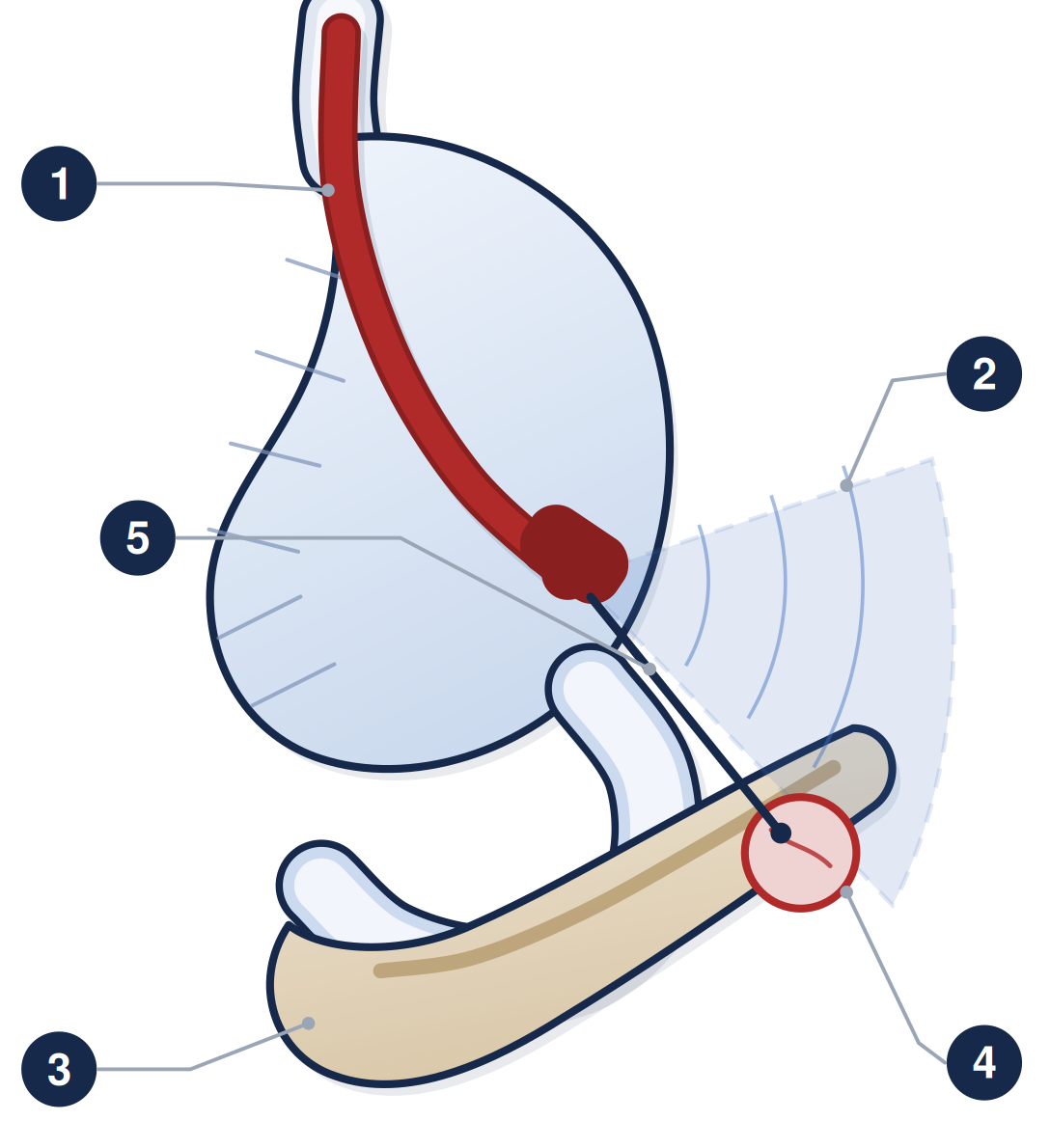
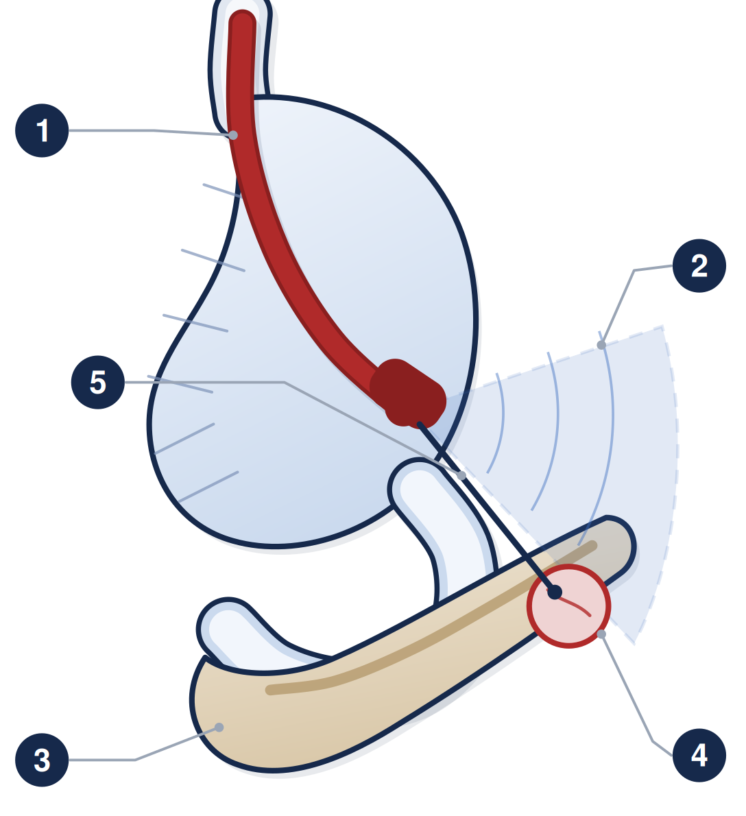
١ المنظار بعد تمريره إلى داخل المعدة ٢ حزمة الموجات فوق الصوتية تعبر جدار المعدة ٣ البنكرياس ٤ المنطقة التي يجري فحصها ٥ الإبرة أثناء أخذ العينة
التحضير
امتنع عن الطعام ست ساعات قبل الفحص، واقتصر على السوائل الصافية حتى ساعتين قبله
تناول أدويتك المعتادة مع رشفة ماء ما لم يُطلب منك غير ذلك
مميعات الدم: الفحص وحده منخفض الخطورة، أما مع الإبرة فهو أعلى خطورة، لذلك اتصل بالوحدة قبل موعدك بسبعة أيام على الأقل. انظر النشرة PE-PROC-16
أخبر وحدة المناظير عن السكري وأمراض القلب والرئة وانقطاع النفس النومي وأي حساسية من المضادات الحيوية وأي مشكلة سابقة مع المهدئات. وإن كنت تستعمل الأنسولين أو أقراص السكري فاتصل بوحدة المناظير أولاً ليُعدَّل الدواء حول فترة الصيام
يوم الإجراء
يُجرى الفحص ببخاخ الحلق ومهدئ عبر الوريد، وأحياناً بتخدير عام. وهو أطول من تنظير المعدة المعتاد، ويستغرق عادةً بين ثلاثين وتسعين دقيقة، لأن المسبار يُحرَّك ببطء عبر المنطقة المطلوب فحصها. وستستلقي على جنبك الأيسر مع مراقبتك طوال الوقت. وإذا أخذت مهدئاً فستحتاج شخصاً بالغاً يوصلك إلى المنزل، وانظر النشرة PE-PROC-17.
ما هي المخاطر؟
الفحص بلا إبرة من أكثر إجراءات التنظير أماناً، أما إضافة الإبرة فتضيف قدراً بسيطاً من الخطورة.
المضاعفة
كم مرة تحدث
ألم الحلق والانتفاخ
شائع ومتوقع لا مضاعفة، ويزول خلال يوم
نزف بعد أخذ عينة بالإبرة
غير شائع، أقل من واحد من كل مئة، ونادراً ما يكون مهماً
التهاب البنكرياس بعد أخذ عينة بالإبرة
نحو واحد من كل مئتين إلى واحد من كل مئة عند أخذ عينة من البنكرياس
العدوى بعد سحب سائل من كيس
نحو واحد من كل مئة أو أقل
انثقاب جدار الأمعاء
نادر جداً، في حدود واحد من كل ألفين أو أقل في الفحص التشخيصي
انتشار خلايا ورمية على مسار الإبرة
نادر، نحو 3 من كل ألف عند أخذ عينة من البنكرياس، وأندر من أن يُعطى له رقم في المواضع الأخرى. ويعتمد على مسار الإبرة: فقد سُجّل بعد أخذ العينة عبر جدار المعدة ولم يُسجّل بعد أخذها عبر الأمعاء الدقيقة. وحيث يمكن الوصول إلى الآفة بالمسارين نختار الأكثر أماناً
بعد الإجراء
ستفيق في القسم لمدة ساعة إلى ساعتين إن أخذت مهدئاً. وإن اكتفيت ببخاخ الحلق فانتظر ساعة قبل الأكل والشرب، ثم اختبر بلعك برشفات من الماء البارد أولاً.
وسيخبرك المنظّر بما شوهد قبل مغادرتك. وإذا أُخذت عينة فإن نتيجتها تأتي من المختبر وتُناقش في موعد العيادة التالي، وسنخبرك بالتاريخ قبل مغادرتك. وقد لا تحتوي عينة الإبرة أحياناً على مادة كافية لإعطاء إجابة فيلزم تكرارها، وهذا ليس أمراً غريباً ولا يعني أن خطأً قد حدث.
اطلب المساعدة العاجلة إذا
أصابك ألم شديد أو متزايد في البطن أو الظهر، فقد يكون التهاب بنكرياس
ارتفعت حرارتك أو أصابتك رعشة
تقيأت دماً أو خرج منك براز أسود قطراني
أصابك ألم شديد في الصدر أو تورم في الرقبة أو صعوبة في التنفس أو البلع
توجّه إلى قسم الطوارئ، وخذ هذه النشرة معك، وأخبر الطاقم أنك خضعت لهذا الفحص وهل أُخذت عينة بالإبرة.
PE-PROC-08 · للمواعيد والاستفسارات، تواصل مع وحدة المناظير عبر الأرقام التي زوّدتك بها. وإذا كانت حالتك شديدة أو كان الوقت خارج أوقات العمل فتوجّه إلى قسم الطوارئ بدلاً من انتظار الرد. تدعم هذه النشرة مناقشتك مع طبيبك وإجراءات الموافقة المستنيرة ولا تحل محلها. الإصدار 1.0 · تاريخ المراجعة أكتوبر 2028.
هذه النشرة معلومات عامة ولا تغني عن مشورة طبيبك المعالج.
زاد صحي، PE-PROC-08
An endoscopic ultrasound is a camera test with an ultrasound probe built into its tip. Because the probe sits inside the gut, right next to the organ being examined, it gives a far more detailed picture than a scan taken from outside the body.
What it is, and why you are having it
A thin flexible tube is passed through the mouth, as in a normal gastroscopy, but this one carries a small ultrasound probe at its end. From inside the stomach and the first part of the small bowel, the probe can see the pancreas, the bile duct, the wall of the gullet and stomach, and the glands and structures immediately around them.
It is used to look at a lump or cyst in the pancreas, to work out how far a cancer has spread before treatment is planned, to look for small stones in the bile duct that a scan may have missed, and to examine a lump growing in the wall of the gullet or stomach.
A fine needle can be passed down the endoscope and through the gut wall to take samples from a lump or gland, or to drain fluid from a cyst. This is called fine needle aspiration or biopsy. It is done under ultrasound guidance so the needle is watched all the way. If fluid is drawn from a cyst, an antibiotic is usually given as well. Not every EUS involves a needle; you will be told beforehand whether one is planned.
1 the camera passed down into the stomach · 2 the ultrasound beam passing through the stomach wall · 3 pancreas · 4 the area being examined · 5 the needle taking a sample
Getting ready
Have nothing to eat for six hours before, and clear fluids only up to two hours before.
Take your usual medicines with a sip of water unless told otherwise.
Blood thinners: EUS on its own is a low-risk procedure, but EUS with a needle is a higher-risk one, so contact the unit at least seven days before your appointment. See sheet PE-PROC-16.
Tell the endoscopy unit about diabetes, heart or lung disease, sleep apnoea, any allergy to antibiotics, and any previous problem with sedation. If you take insulin or tablets for diabetes, telephone the endoscopy unit first so the dose can be adjusted around the fast.
On the day
EUS is done with throat spray and sedation through a drip, and occasionally under a general anaesthetic. It takes longer than a normal gastroscopy — usually somewhere between 30 and 90 minutes — because the probe has to be worked slowly across the area of interest. You will lie on your left side and be monitored throughout. If you have sedation you will need a responsible adult to take you home; see sheet PE-PROC-17.
What are the risks?
EUS without a needle is one of the safest endoscopic procedures. Adding a needle adds a small amount of risk.
Complication
How often it happens
Sore throat and bloating
Common, and expected rather than a complication. Settles within a day.
Bleeding after a needle sample
Uncommon — fewer than 1 in 100, and rarely significant.
Pancreatitis after a needle sample
Around 1 in 200 to 1 in 100 when the pancreas is sampled.
Infection after draining a cyst
Around 1 in 100 or fewer.
Perforation — a tear in the gut wall
Very rare, in the region of 1 in 2,000 or fewer for a diagnostic examination.
Cancer cells spreading along the needle track
Rare — about 3 in every 1,000 where the pancreas is sampled, and too rare elsewhere to put a number to. It depends on the route the needle takes: it has been reported after sampling through the stomach wall and not after sampling through the small bowel. Where a lesion can be reached either way, we take the safer route.
Afterwards
You will recover in the department for one to two hours if you had sedation. If you had throat spray, wait an hour before eating or drinking, then test your swallow with a few sips of cold water first.
The endoscopist will tell you what was seen before you leave. If a sample was taken, the result comes from the laboratory and is discussed at your next clinic appointment — we will tell you the date before you go home. A needle sample sometimes does not contain enough material to give an answer, and has to be repeated; this is not unusual and does not mean anything has gone wrong.
Get urgent help if
You have severe or worsening abdominal or back pain — this may be pancreatitis.
You have a fever or shivering.
You vomit blood, or pass black tarry stools.
You have severe chest pain, swelling in the neck, or difficulty breathing or swallowing.
Go to the Emergency Department, take this sheet with you, and tell the staff you have had an endoscopic ultrasound and whether a needle sample was taken.
PE-PROC-08 · For appointments and questions, contact the endoscopy unit using the numbers it has given you. If you are seriously unwell, or it is outside working hours, go to the Emergency Department rather than waiting for a reply. This sheet supports the discussion with your doctor and the consent process; it does not replace them. Version 1.0 · review date October 2028.
This leaflet is general information and does not replace advice from your own doctor.