PE-PROC-07الإصدار 1.0 · آخر مراجعة أكتوبر 2026 · المراجعة القادمة أكتوبر 2028Version 1.0 · Last reviewed October 2026 · Next review October 2028
إجراء يستخدم الكاميرا والأشعة السينية للوصول إلى القناة الصفراوية وقناة البنكرياس ولعلاج مشاكلهما. وهو في الممارسة الحديثة علاج لا فحص تشخيصي، إذ يُشخَّص المرض بالتصوير أولاً. يُرجى قراءة هذه النشرة قبل موعدك.
ما هو الإجراء ولماذا تحتاجه
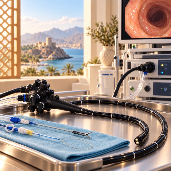
يُدخل منظار خاص ذو رؤية جانبية عبر الفم حتى الموضع في الأمعاء الدقيقة الذي تنفتح فيه القناة الصفراوية وقناة البنكرياس. ثم يُمرَّر أنبوب رفيع داخل القناة وتُحقن صبغة الأشعة، وتُعالَج المشكلة عبر المنظار بتوجيه الأشعة. ولا يُجرى أي شق في خارج الجسم.
وأكثر الأسباب شيوعاً حصوات القناة الصفراوية، والانسداد المسبب لليرقان، والتهاب القنوات الصفراوية، وتسرب العصارة الصفراوية بعد استئصال المرارة، وتغيير الدعامة أو إزالتها. وللوصول إلى القناة تُقطع عادةً العضلة الصغيرة عند فتحتها بتيار كهربائي، أو تُوسَّع ببالون. ثم تُسحب الحصوات بسلة أو بالون، ويُبقى التضيّق مفتوحاً بأنبوب صغير يُسمى الدعامة.
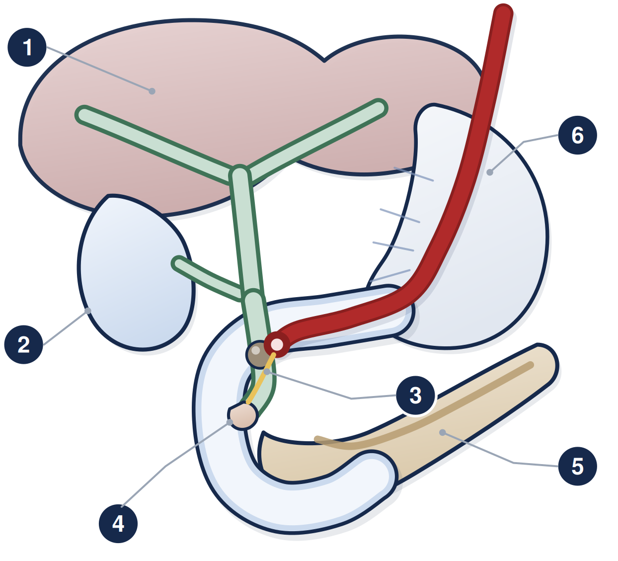
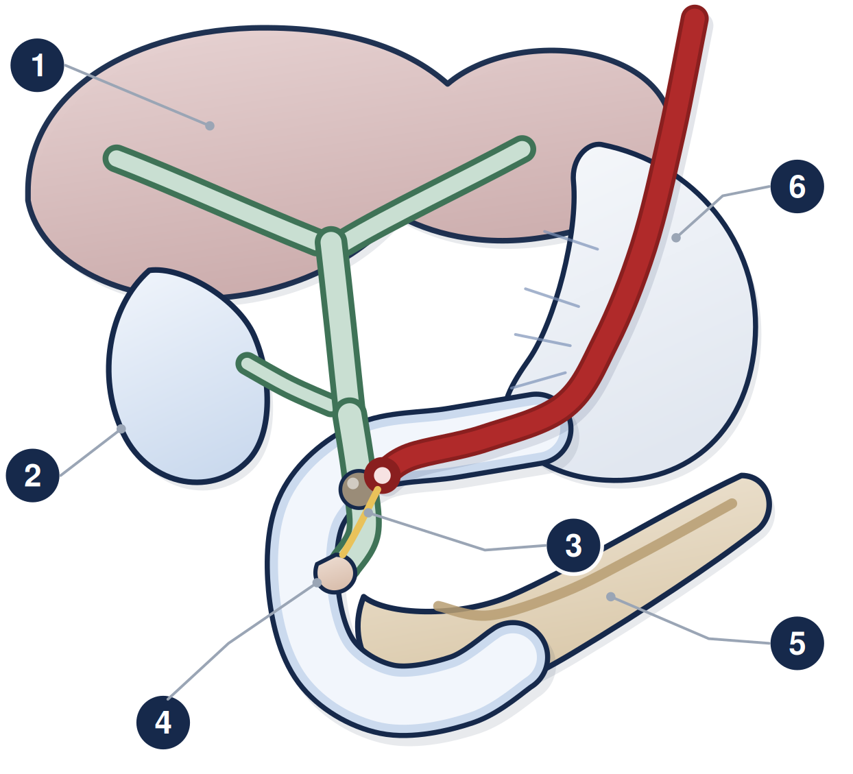
١ الكبد والقنوات الصفراوية داخله ٢ المرارة ٣ القناة الصفراوية وفيها حصاة ٤ فتحة تصريف القناة الصفراوية وقناة البنكرياس في الاثني عشر ٥ البنكرياس وقناته ٦ المنظار جانبي الرؤية داخل الاثني عشر
التحضير
امتنع عن الطعام ست ساعات قبل الإجراء، واقتصر على السوائل الصافية حتى ساعتين قبله
أخبر وحدة المناظير إن سبق أن تحسّست من صبغة الأشعة، وإن كان هناك احتمال للحمل
مميعات الدم مهمة هنا. فالإجراء مع قطع العضلة أعلى خطورة للنزف، لذلك يجب إيقاف بعض الأدوية ولعدد محدد من الأيام. اتصل بالوحدة قبل موعدك بسبعة أيام على الأقل واتبع الخطة المكتوبة التي نعطيك إياها، وانظر النشرة PE-PROC-16. ولا توقف مميع الدم من تلقاء نفسك أبداً
لا تُعطى المضادات الحيوية بشكل روتيني، لكنها تُعطى إذا توقعنا أن التصريف لن يكتمل، أو إذا كانت مناعتك مثبطة بشدة، أو في بعض الإجراءات المتخصصة
أخبر وحدة المناظير عن السكري وأمراض القلب والرئة وانقطاع النفس النومي وأي مشكلة سابقة مع المهدئات أو التخدير. وإن كنت تستعمل الأنسولين أو أقراص السكري فاتصل بوحدة المناظير أولاً ليُعدَّل الدواء حول فترة الصيام
يوم الإجراء
يُجرى الإجراء بمهدئ عبر الوريد، أو بتخدير عام في الحالات الأطول أو الأعقد ولمن لا يستطيع البقاء ساكناً. ويستغرق نحو ثلاثين دقيقة عادةً، وقد يمتد من ربع ساعة إلى ساعة ونصف. وستُوضع في الوضعية التي يطلبها المنظّر، غالباً على البطن أو على الجانب الأيسر، مع مراقبتك طوال الوقت.
ويُعطى كثير من المرضى تحميلة شرجية من دواء مضاد للالتهاب (ديكلوفيناك أو إندوميثاسين) قبل الإجراء مباشرة أو أثناءه. وهي ليست للألم، بل تقلل كثيراً احتمال التهاب البنكرياس بعده، وهو أكثر مضاعفات الإجراء شيوعاً. وتُعطى قبل الإجراء مباشرة. أخبري وحدة المناظير إن كنتِ حاملاً أو يحتمل أن تكوني كذلك، أو كانت لديك قرحة معدة حديثة أو ضعف في وظائف الكلى، أو سبق أن تحسّست من دواء مضاد للالتهاب. وأخبري وحدة المناظير عن الحمل على كل حال، لأن الأشعة السينية تُستخدم طوال هذا الإجراء.
ما هي المخاطر؟
يحمل هذا الإجراء خطورة أعلى من تنظير المعدة أو القولون المعتاد، ولهذا لا يُجرى إلا حين تكون الفائدة واضحة. ويصاب نحو واحد من كل خمسة عشر مريضاً بمضاعفة ما، ومعظمها خفيف ويهدأ بالبقاء ليلة أو ليلتين في المستشفى.
المضاعفة
كم مرة تحدث
التهاب البنكرياس
الأكثر شيوعاً. نحو 3 إلى 9 من كل مئة مريض، ونحو واحد من كل سبعة في الفئات الأعلى خطورة. يسبب ألماً شديداً ويستدعي عادةً البقاء في المستشفى
نزف من موضع قطع العضلة
نحو واحد من كل مئة. يتوقف عادةً من تلقاء نفسه أو يُسيطَر عليه أثناء الإجراء
التهاب القنوات الصفراوية
نحو 1 إلى 3 من كل مئة، ويزداد احتماله إذا تعذّر تصريف القناة تماماً
انثقاب جدار الأمعاء أو القناة
نحو 6 من كل ألف. غير شائع لكنه خطير وقد يستدعي الجراحة
اضطراب التنفس أو النبض أو ضغط الدم أثناء التخدير
الانخفاضات العابرة شائعة وتُعالَج فوراً، أما الأحداث الخطيرة فنادرة
الوفاة
نادرة، نحو 3 من كل ألف، ومعظمها لدى مرضى شديدي المرض أصلاً
بعد الإجراء
ستُراقب في منطقة الإفاقة، ويبقى معظم المرضى في المستشفى بقية اليوم، وبعضهم يبيت ليلة. وسيخبرك الفريق متى يمكنك الأكل والشرب. وألم الحلق البسيط والانتفاخ أمران طبيعيان. ولأنك أخذت مهدئاً أو تخديراً عاماً فلا بد من شخص بالغ مسؤول يوصلك إلى المنزل ويبقى معك، ولا يجوز لك القيادة أو تشغيل الآلات أو تناول الكحول أو توقيع أي أمر مهم لمدة 24 ساعة، وانظر النشرة PE-PROC-17.
وثمة أمران آخران يحسن معرفتهما مسبقاً. فأحياناً يتعذر دخول القناة رغم محاولة صحيحة، فيُرتَّب حينها تنظير ثانٍ أو تصريف بطريق آخر. وأحياناً تُترك دعامة بلاستيكية رفيعة في قناة البنكرياس لحمايتها، وهي تخرج من تلقاء نفسها عادةً، وتؤكد صورة أشعة بسيطة بعد أسبوع أو أسبوعين أنها خرجت.
إذا وُضعت لك دعامة بلاستيكية — هذا الجزء مهم
الدعامة البلاستيكية أنبوب مؤقت، ويجب إزالتها أو تغييرها، عادةً عند نحو ثلاثة أشهر وبحد أقصى ستة. فإذا تُركت في مكانها فستنسد في النهاية، والدعامة المسدودة قد تسبب التهاباً خطيراً في القناة الصفراوية بسرعة، وأحياناً خلال يوم واحد. فتأكد من مغادرتك ومعك تاريخ مكتوب للإجراء التالي، واتصل بوحدة المناظير إن لم يصلك ذلك الموعد.
اطلب المساعدة العاجلة إذا
أصابك ألم بطني شديد أو متزايد، وبخاصة إن كان ينفذ إلى الظهر، فقد يكون التهاب بنكرياس
ارتفعت حرارتك أو أصابتك رعشة وقشعريرة، فقد يكون التهاباً في القناة الصفراوية أو دعامة مسدودة
عاد اصفرار العينين أو الجلد، أو صار بولك داكناً، أو صار برازك شاحب اللون
تقيأت دماً أو خرج منك براز أسود قطراني
أصابك ألم في الصدر أو صعوبة في التنفس
لا تنتظر موعدك التالي. توجّه إلى قسم الطوارئ، وخذ هذه النشرة معك، وأخبر الطاقم أنك خضعت لهذا الإجراء وهل وُضعت لك دعامة.
PE-PROC-07 · للمواعيد والاستفسارات، تواصل مع وحدة المناظير عبر الأرقام التي زوّدتك بها. وإذا كانت حالتك شديدة أو كان الوقت خارج أوقات العمل فتوجّه إلى قسم الطوارئ بدلاً من انتظار الرد. تدعم هذه النشرة مناقشتك مع طبيبك وإجراءات الموافقة المستنيرة ولا تحل محلها. الإصدار 1.0 · تاريخ المراجعة أكتوبر 2028.
هذه النشرة معلومات عامة ولا تغني عن مشورة طبيبك المعالج.
زاد صحي، PE-PROC-07
ERCP is a procedure that uses a camera and X-rays to reach the bile duct and the pancreatic duct, and to treat problems in them. In modern practice it is a treatment rather than a test — scans are used to make the diagnosis first. Please read this before your appointment.
What it is, and why you are having it
A special endoscope that looks sideways is passed through the mouth to the point in the small bowel where the bile duct and pancreatic duct open. A fine tube is threaded into the duct, X-ray dye is injected, and the problem is treated through the endoscope under X-ray guidance. Nothing is cut on the outside of your body.
The usual reasons are stones in the bile duct, a blockage causing jaundice, infection in the bile (cholangitis), a leak of bile after gallbladder surgery, and changing or removing a stent. To reach the duct, the small muscle at its opening is usually cut with an electric current (a sphincterotomy) or stretched with a balloon. Stones are then pulled out with a basket or balloon, and a narrowing is held open with a small tube called a stent.
1 the liver and the ducts inside it · 2 gallbladder · 3 the bile duct, with a stone in it · 4 where the ducts drain into the bowel · 5 pancreas and its duct · 6 the side-viewing camera
Getting ready
Have nothing to eat for six hours before, and clear fluids only up to two hours before.
Tell the endoscopy unit if you have ever reacted to X-ray dye, and if there is any chance you are pregnant.
Blood thinners matter here. ERCP with a sphincterotomy is a higher-bleeding-risk procedure, so some medicines have to be stopped, and for a set number of days. Contact the unit at least seven days before your appointment and follow the written plan we give you — see sheet PE-PROC-16. Never stop a blood thinner on your own.
Antibiotics are not given routinely, but they are given if we expect the drainage to be incomplete, if your immune system is severely suppressed, or for certain specialised procedures.
Tell the endoscopy unit about diabetes, heart or lung disease, sleep apnoea, and any previous problem with sedation or anaesthetic. If you take insulin or tablets for diabetes, telephone the endoscopy unit first so the dose can be adjusted around the fast.
On the day
ERCP is done with sedation through a drip, or under a general anaesthetic for longer or more complex cases and for people who cannot lie still. The procedure usually takes about 30 minutes, and can take anywhere from 15 minutes to an hour and a half. You will be positioned as the endoscopist asks, usually lying on your front or on your left side, and monitored throughout.
Many patients are given a suppository of an anti-inflammatory medicine (diclofenac or indomethacin) into the back passage just before or during the procedure. This is not for pain — it substantially reduces the chance of pancreatitis afterwards, which is the commonest complication of ERCP. It is given immediately before the procedure. Tell the endoscopy unit if you are pregnant or might be, have had a stomach ulcer recently, have poor kidney function, or have ever reacted badly to an anti-inflammatory medicine. Tell the endoscopy unit about pregnancy in any case, because X-rays are used throughout this procedure.
What are the risks?
ERCP carries more risk than a standard gastroscopy or colonoscopy, which is why it is only done when the benefit is clear. About 1 in 15 people have some complication; most are mild and settle with a night or two in hospital.
Complication
How often it happens
Pancreatitis — inflammation of the pancreas
The commonest. Roughly 3 to 9 in every 100 people, and around 1 in 7 if you are in a higher-risk group. It causes severe pain and usually needs a hospital stay.
Bleeding, from the cut at the duct opening
Around 1 in 100. Usually stops on its own or is controlled at the time.
Infection in the bile (cholangitis)
Around 1 to 3 in 100, and more likely if the duct cannot be fully drained.
Perforation — a tear in the bowel or duct wall
About 6 in every 1,000. Uncommon but serious, and may need surgery.
Problems with breathing, heart rate or blood pressure during sedation
Short-lived dips are common and treated on the spot; serious events are rare.
Death
Rare — about 3 in every 1,000, mostly in people who are already seriously ill.
Afterwards
You will be watched in the recovery area, and most people stay in hospital for the rest of the day; some stay overnight. The team will tell you when you can eat and drink. A sore throat and some bloating are normal. Because you have had sedation or an anaesthetic, a responsible adult must take you home and stay with you, and you must not drive, use machinery, drink alcohol or sign anything important for 24 hours — see sheet PE-PROC-17.
Two other things are worth knowing in advance. Occasionally the duct cannot be entered despite a proper attempt; a second ERCP, or drainage by a different route, is then arranged. And a fine plastic stent is sometimes left in the pancreatic duct to protect it — it usually passes on its own, and a plain X-ray a week or two later confirms that it has gone.
If you have been given a plastic stent — this part is important
A plastic stent is a temporary tube. It must be removed or exchanged, normally at about three months and at the latest by six. If it is left in place it will eventually block, and a blocked stent can cause a serious infection of the bile duct quickly — sometimes within a day. Make sure you leave with a date for your next procedure written down, and contact the endoscopy unit if that appointment does not arrive.
Get urgent help if
You have severe or worsening abdominal pain, especially boring through to the back — this may be pancreatitis.
You have a fever or shivering and shaking chills — this may be infection in the bile, or a blocked stent.
Yellowing of the eyes or skin returns, or your urine goes dark, or your stools become pale.
You vomit blood, or pass black tarry stools.
You have chest pain or difficulty breathing.
Do not wait for your next appointment. Go to the Emergency Department, take this sheet with you, and tell the staff you have had an ERCP and whether a stent was placed.
PE-PROC-07 · For appointments and questions, contact the endoscopy unit using the numbers it has given you. If you are seriously unwell, or it is outside working hours, go to the Emergency Department rather than waiting for a reply. This sheet supports the discussion with your doctor and the consent process; it does not replace them. Version 1.0 · review date October 2028.
This leaflet is general information and does not replace advice from your own doctor.