Having a feeding tube placed into the stomach (PEG)
ما تحتاج معرفته قبل الفحص وبعده
What you need to know before and after the test
PE-PROC-09الإصدار 1.0 · آخر مراجعة أكتوبر 2026 · المراجعة القادمة أكتوبر 2028Version 1.0 · Last reviewed October 2026 · Next review October 2028
أنبوب تغذية لين يمر عبر جلد البطن إلى المعدة مباشرة، ويُركَّب بالمنظار فلا يحتاج شقاً أو عملية مفتوحة. هذه النشرة للمريض ولمن سيرعى الأنبوب من الأهل أو مقدمي الرعاية.
ما هو الإجراء ولماذا تحتاجه
يُستخدم هذا الأنبوب حين يكون البلع غير آمن أو غير كافٍ للحفاظ على التغذية، ويُتوقع أن يستمر ذلك أكثر من نحو أربعة أسابيع. وأكثر الأسباب شيوعاً السكتة الدماغية، والأمراض العصبية التقدمية مثل مرض العصبون الحركي، وسرطانات الرأس والعنق والمريء، وغالباً أثناء العلاج الإشعاعي.
ووجود الأنبوب لا يمنعك من تذوق الطعام أو الأكل للمتعة إن كان ذلك آمناً لك. فالغرض منه ضمان وصول الغذاء والسوائل والأدوية بصورة موثوقة، حتى لا يتعطل العلاج والتعافي بسبب نقص الوزن والجفاف.
الجانب الصريح
تركيب الأنبوب إجراء صغير، لكنه يُقدَّم لمرضى شديدي المرض أصلاً، وهذا ما يحدد المآل لا الأنبوب نفسه. وفي دراسة وطنية كبيرة تُوفي نحو واحد من كل عشرين مريضاً خلال ثلاثين يوماً من التركيب، بسبب المرض الأساسي في الغالب الأعم لا بسبب الإجراء. وقد انخفض هذا الرقم إلى أقل من نصفه خلال الخمس عشرة سنة الماضية بفضل الانتقاء الأدق للمرضى. ولهذا يُتخذ القرار بعناية معك ومع أسرتك، ولهذا لا يُنصح بالأنبوب في الخرف المتقدم أو حين يكون العمر المتوقع قصيراً جداً. اطلب منا أن نشرح لك ما نتوقع أن يحققه الأنبوب في حالتك أنت.
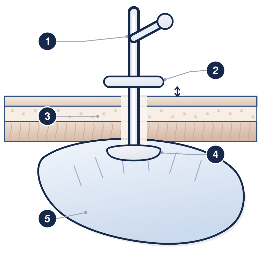
١ أنبوب التغذية ومنفذ الإطعام ٢ الصفيحة الخارجية وتبعد عن الجلد بمقدار عرض طرف الإصبع ٣ طبقات جدار البطن ٤ القرص الداخلي الليّن داخل المعدة ٥ المعدة
التحضير
الامتناع عن الطعام ست ساعات قبل الإجراء، والسوائل الصافية حتى ساعتين قبله، ومدة أطول إذا كانت المعدة تفرغ ببطء
تُعطى جرعة واحدة من مضاد حيوي في الوريد قبل الإجراء بنحو ثلاثين دقيقة، وهذا إجراء معتاد يقلل كثيراً احتمال العدوى في موضع الأنبوب
مميعات الدم: تركيب الأنبوب إجراء أعلى خطورة للنزف، لذلك اتصل بالوحدة قبل الموعد بسبعة أيام على الأقل. انظر النشرة PE-PROC-16
تُناقش الموافقة معك إن كنت قادراً على المشاركة في القرار، وإن لم تكن فيُتخذ القرار بما فيه مصلحتك مع أسرتك والفريق الطبي
يوم الإجراء
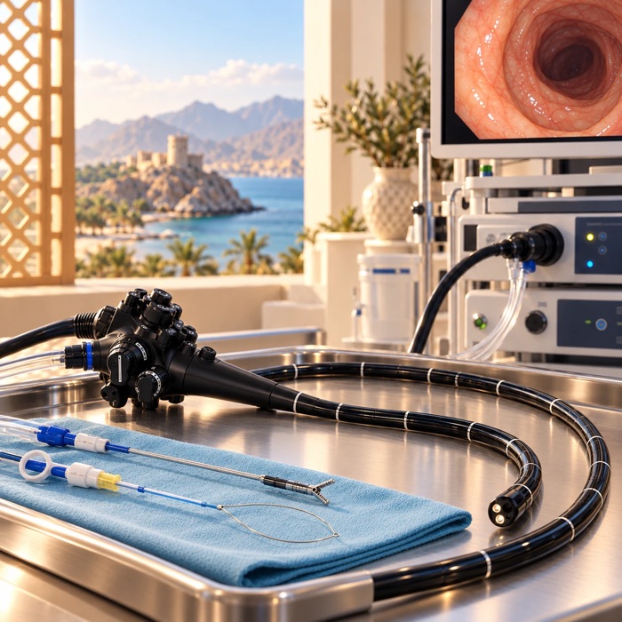
يُجرى التركيب بمهدئ عبر الوريد. يُدخل المنظار إلى المعدة ويُستخدم الهواء ليقرّب جدار المعدة من جدار البطن. ويُحدَّد الموضع المناسب على الجلد بالضغط على البطن وبرؤية ضوء المنظار عبر الجلد. ثم يُحقن مخدر موضعي ويُعمل ثقب صغير ويُمرَّر خيط إلى داخل المعدة يلتقطه المنظار ويُخرجه من الفم. ويُربط أنبوب التغذية بالخيط ويُسحب عبر المريء ليخرج من جدار البطن. ويثبته قرص لين داخل المعدة، وصفيحة صغيرة على الجلد تحفظ وضعه.
وتبدأ التغذية عادةً خلال ثلاث إلى أربع ساعات من التركيب غير المعقّد. أما النصيحة القديمة بالصيام طوال الليل بعده فلم تعد مستخدمة.
ما هي المخاطر؟
المضاعفة
كم مرة تحدث
العدوى حول موضع الأنبوب
الأكثر شيوعاً، أقل من واحد من كل عشرة، وتقل بفضل المضاد الحيوي المعطى مسبقاً. وتُعالَج عادةً بالمضادات الحيوية
النزف
نحو 1 إلى 2 من كل مئة
التهاب الغشاء البريتوني داخل البطن
نحو واحد من كل مئة. خطير ويحتاج علاجاً عاجلاً
انثقاب القولون، وقد يقع جزء منه أمام المعدة
نادر، نحو اثنين من كل ألف
انغراس القرص الداخلي في جدار المعدة
نحو 1 إلى 4 من كل مئة مع الوقت، ويُمنع بإبقاء الصفيحة الجلدية بعيدة عن الجلد بمقدار طرف الإصبع بعد التئام الموضع، وبتحريك الأنبوب يومياً كما يُشرح لك، وانظر النشرة PE-PROC-10
دخول الطعام أو السوائل إلى الرئتين
خطر لدى من لا يستطيع حماية مجرى تنفسه. والأنبوب يقلل ما يجب بلعه لكنه لا يمنع التهاب الرئة الناتج عن الشفط، ومن المهم ألا يُتوقع منه ذلك
نمو نسيج زائد حول الموضع
شائع وغير ضار عادةً، لكن أخبر الممرض إن نزف أو أفرز سائلاً
بعد الإجراء
ستُراقَب بضع ساعات ويُتحقق من وضع الأنبوب وموضع الجلد. وتُزاد التغذية تدريجياً بإشراف أخصائي التغذية. والألم حول الموضع في اليوم الأول أو الثاني أمر طبيعي ويهدأ بمسكنات بسيطة.
ولا يجوز نزع الأنبوب قبل مرور أربعة أسابيع على الأقل، لأن القناة بين المعدة والجلد تحتاج هذه المدة لتتكوّن. أما تفاصيل التغذية والغسل والتنظيف وما يجب عمله إن انسد الأنبوب أو خرج فترد في النشرة PE-PROC-10، فاقرأها قبل مغادرتك واحرص على أن يقرأها من سيساعدك في المنزل.
اطلب المساعدة العاجلة إذا
خرج الأنبوب. هذا أمر عاجل في أي وقت، فقد تبدأ الفتحة بالانغلاق خلال ساعات. غطِّ الموضع بضمادة نظيفة جافة، واحضر ومعك الأنبوب الذي خرج، وتوجّه إلى المستشفى فوراً. لا تحاول إعادته بنفسك ولا تُدخل شيئاً في الفتحة
أصابك ألم بطني شديد، أو صار بطنك متصلباً أو منتفخاً، أو ازداد الألم بدلاً من أن يخف
ارتفعت حرارتك أو أصابتك رعشة
ظهر احمرار متسع أو تورم أو سخونة أو صديد حول موضع الأنبوب
تسرب الغذاء أو محتويات المعدة حول الأنبوب من الخارج
أصابك ضيق في التنفس أو سعال أثناء التغذية أو بعدها
PE-PROC-09 · للمواعيد والاستفسارات، تواصل مع وحدة المناظير عبر الأرقام التي زوّدتك بها. وإذا كانت حالتك شديدة أو كان الوقت خارج أوقات العمل فتوجّه إلى قسم الطوارئ بدلاً من انتظار الرد. تدعم هذه النشرة مناقشتك مع طبيبك وإجراءات الموافقة المستنيرة ولا تحل محلها. الإصدار 1.0 · تاريخ المراجعة أكتوبر 2028.
هذه النشرة معلومات عامة ولا تغني عن مشورة طبيبك المعالج.
زاد صحي، PE-PROC-09
A PEG is a soft feeding tube that passes through the skin of the abdomen directly into the stomach. It is placed using an endoscope, so no cut or open operation is needed. This sheet is for the patient and for the family or carer who will look after the tube.
What it is, and why you are having it
A PEG is used when swallowing is unsafe or not enough to maintain nutrition, and this is expected to last more than about four weeks. The commonest reasons are a stroke, a progressive neurological illness such as motor neurone disease, and cancer of the head, neck or gullet — often during radiotherapy.
A PEG does not stop you tasting food or eating for pleasure if it is safe for you to do so. It is there to make sure that nutrition, fluid and medicines get in reliably, so that treatment and recovery are not held back by weight loss and dehydration.
The honest part
A PEG is a small procedure, but it is offered to people who are already seriously unwell, and that is what drives the outlook rather than the tube itself. In a large national study, about 1 in 20 people died within 30 days of having a PEG placed — almost always from the underlying illness rather than from the procedure. That figure has fallen by more than half over the past fifteen years, because patients are now selected more carefully. This is why the decision is made carefully, with you and your family, and why a PEG is not recommended in advanced dementia or when life expectancy is very short. Ask the endoscopy unit to explain what a PEG is expected to achieve in your own situation.
1 the feeding tube and its port · 2 the outer plate, a fingertip's width clear of the skin · 3 the abdominal wall · 4 the soft internal disc inside the stomach · 5 the stomach
Getting ready
Nothing to eat for six hours before, and clear fluids only up to two hours before. Longer if the stomach empties slowly.
A single dose of antibiotic is given into a vein about 30 minutes before the procedure. This is standard and significantly reduces the chance of infection at the site.
Blood thinners: PEG placement is a higher-bleeding-risk procedure, so contact the unit at least seven days before. See sheet PE-PROC-16.
Consent is discussed with you if you are able to take part in the decision. If you are not, the decision is made in your best interests with your family and the medical team.
On the day
The procedure is done with sedation through a drip. An endoscope is passed into the stomach and air is used to bring the stomach wall against the abdominal wall. The right spot on the skin is found by pressing on the abdomen and by seeing the endoscope light through the skin. Local anaesthetic is injected, a small nick is made, and a thread is passed through into the stomach, caught by the endoscope and brought out through the mouth. The feeding tube is tied to the thread and drawn down the gullet and out through the abdominal wall. A soft internal disc holds it inside the stomach and a small plate on the skin holds it in position.
Feeding usually starts within three to four hours of an uncomplicated placement. The old advice to fast overnight afterwards is no longer used.
What are the risks?
Complication
How often it happens
Infection around the tube site
The commonest — fewer than 1 in 10 people, and lower because of the antibiotic given beforehand. Usually treated with antibiotics.
Bleeding
Around 1 to 2 in every 100.
Peritonitis — infection inside the abdomen
Around 1 in every 100. Serious, and needs urgent treatment.
Perforation of the colon, part of which can lie in front of the stomach
Rare — about 2 in every 1,000.
Buried bumper — the internal disc grows into the stomach wall
Around 1 to 4 in every 100 over time. Prevented by holding the skin plate about a fingertip clear of the skin once the site has healed, and by moving the tube daily as instructed — see sheet PE-PROC-10.
Food or fluid going into the lungs
A risk in people who cannot protect their airway. A PEG reduces the amount that has to be swallowed, but it does not prevent chest infection from aspiration, and it is important not to expect that of it.
Overgrowth of tissue at the site
Common and usually harmless, but tell the nurse if it bleeds or weeps.
Afterwards
You will be watched for a few hours and the tube position and the skin site checked. Feeding is built up gradually by the dietitian. Pain around the site for the first day or two is normal and settles with simple painkillers.
The tube must not be removed for at least four weeks, because the channel between the stomach and the skin takes that long to form. Detailed instructions on feeding, flushing, cleaning and what to do if the tube blocks or comes out are in sheet PE-PROC-10 — please read it before you go home, and make sure whoever helps you at home reads it too.
Get urgent help if
The tube falls out. This is urgent at any time — the opening can start to close within a few hours. Cover it with a clean dry pad, bring the tube with you, and come to hospital straight away. Do not try to push it back in and do not put anything down the opening.
You have severe abdominal pain, a hard or swollen abdomen, or pain that gets worse rather than better.
You have a fever or shivering.
There is spreading redness, swelling, heat or pus around the tube site.
Feed or stomach contents are leaking around the outside of the tube.
You are breathless or coughing during or after feeds.
PE-PROC-09 · For appointments and questions, contact the endoscopy unit using the numbers it has given you. If you are seriously unwell, or it is outside working hours, go to the Emergency Department rather than waiting for a reply. This sheet supports the discussion with your doctor and the consent process; it does not replace them. Version 1.0 · review date October 2028.
This leaflet is general information and does not replace advice from your own doctor.