PE-PROC-06الإصدار 1.0 · آخر مراجعة أكتوبر 2026 · المراجعة القادمة أكتوبر 2028Version 1.0 · Last reviewed October 2026 · Next review October 2028
تنظير الجيب فحص قصير بالكاميرا للجيب الداخلي الذي صُنع من أمعائك الدقيقة عند استئصال الأمعاء الغليظة. يُرجى قراءة هذه النشرة قبل موعدك وإحضارها معك.
ما هو الإجراء ولماذا تحتاجه
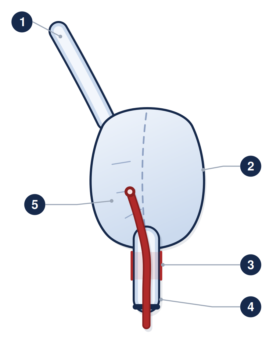
عند استئصال القولون والمستقيم يمكن طيّ نهاية الأمعاء الدقيقة لتكوين خزّان يُسمى الجيب، ويُوصل بالشرج ليتم التبرز بالطريقة الطبيعية. وعادةً يُترك جزء قصير من المستقيم عند موضع الوصل يُسمى الحافة. ويستخدم التنظير كاميرا رفيعة مرنة تُدخل عبر الشرج لفحص الجيب والحافة والأمعاء الدقيقة أعلى الجيب مباشرة.
ويُجرى لسببين. الأول تحديد سبب الأعراض، مثل زيادة عدد مرات التبرز أو الإلحاح أو النزف أو الألم أو التسرب، وقد يكون السبب التهاب الجيب أو التهاب الحافة أو تضيّقاً عند موضع الوصل أو مرضاً شبيهاً بداء كرون في الجيب. والثاني المتابعة الدورية بأخذ عينات صغيرة للبحث عن تغيرات ما قبل سرطانية مبكرة.
وتعتمد وتيرة تنظير المتابعة على حالتك أنت، وبخاصة ما إذا وُجد تغير ما قبل سرطاني أو سرطان في الأمعاء التي استُؤصلت، وما إذا كان لديك أيضاً التهاب الأقنية الصفراوية المصلب الأولي أو التهاب مزمن في الجيب. فبعض المرضى يحتاجونه سنوياً وآخرون كل بضع سنوات، وسيكتب فريقك الفترة الخاصة بك على هذه النشرة. وسرطان الجيب نادر، أقل من واحد من كل مئة. وما تبحث عنه المتابعة فعلياً هو التغير ما قبل السرطاني الأبكر، ويظهر لدى نحو اثنين من كل مئة شخص خلال خمس عشرة سنة وهو قابل للعلاج عند اكتشافه. وكلاهما أقل احتمالاً بكثير إن لم يوجد تغير ما قبل سرطاني في عمليتك الأصلية.
١ نهاية الأمعاء الدقيقة الداخلة إلى الجيب ٢ الجيب المصنوع بطيّ الأمعاء على نفسها ٣ حافة المستقيم القصيرة المتبقية عند موضع الوصل ٤ القناة الشرجية ٥ الكاميرا الداخلة عبر الشرج
التحضير
لا تحتاج إلى التنظيف الكامل للأمعاء المستخدم في تنظير القولون، فالحقنة الشرجية تكفي عادةً، وستحدد لك رسالة الموعد وقت استعمالها، وغالباً قبل نحو ثلاث ساعات من مغادرتك المنزل
امتنع عن الطعام ست ساعات قبل الفحص، واقتصر على السوائل الصافية حتى ساعتين قبله
تناول أدويتك المعتادة ما لم يُطلب منك غير ذلك. وإن كنت تستعمل الأنسولين أو أقراص السكري فاتصل بوحدة المناظير أولاً، فالجرعة تحتاج تعديلاً عادةً حول فترة الصيام
مميعات الدم: لا يستدعي التنظير التشخيصي مع الخزعات إيقافها عادةً، لكن أخبر وحدة المناظير بما تتناوله. وإذا رُجّح أن يحتاج تضيّق إلى توسيع فقد تتغير الخطة، وانظر النشرة PE-PROC-16
يوم الإجراء
يتحمل معظم المرضى هذا الفحص بلا أي تخدير. فهو قصير، نحو عشر دقائق عادةً، والجيب قريب من الشرج فلا تقطع الكاميرا مسافة طويلة. ويمكنك اختيار غاز الاستنشاق أو مهدئ عبر الوريد إن فضّلت ذلك. وإذا أخذت مهدئاً فستحتاج شخصاً بالغاً يوصلك إلى المنزل، ولا يجوز لك القيادة لمدة 24 ساعة، وانظر النشرة PE-PROC-17.
ستستلقي على جنبك الأيسر. ويُستخدم الهواء أو ثاني أكسيد الكربون لفتح الجيب ليُرى جيداً، وتُؤخذ عادةً عينات نسيجية صغيرة غير مؤلمة من الأمعاء الدقيقة أعلى الجيب ومن الجيب نفسه ومن حافة المستقيم. وعينات الحافة مهمة، لأن هذا الجزء المتبقي من المستقيم هو الموضع الأرجح لظهور التغير ما قبل السرطاني. وتُؤخذ العينات أعلى الجزء الحساس من القناة الشرجية، ولهذا لا تسبب ألماً.
ما هي المخاطر؟
تنظير الجيب فحص قصير منخفض الخطورة ومضاعفاته الخطيرة نادرة جداً. ولا يوجد رقم منشور لمعدل مضاعفات تنظير الجيب تحديداً، لذلك نذكر مخاطر التنظير السيني المرن وهو فحص مشابه: النزف بعد الخزعة وانثقاب جدار الأمعاء كلاهما نادر، في حدود واحد من كل ألف أو أقل. أما الانتفاخ والغازات بعد الفحص فهي أمور متوقعة لا مضاعفات. وإذا استُخدم التخدير فتنطبق مخاطره الخاصة، وانظر النشرة PE-PROC-17.
وإذا وُسّع تضيّق عند موضع الوصل أثناء الفحص فإن ذلك يحمل خطورة أعلى وسيُشرح لك وتُؤخذ موافقتك عليه بصورة منفصلة.
بعد الإجراء
يمكنك الأكل والشرب فوراً ما لم يُطلب منك غير ذلك، والعودة إلى نشاطك المعتاد في اليوم نفسه إن لم تأخذ مهدئاً. والانتفاخ والغازات وقليل من الدم عند المسح أمور شائعة ليوم أو يومين.
وسيشرح لك المنظّر ما شوهد قبل مغادرتك. أما نتائج الخزعات فتستغرق أسبوعين إلى أربعة وتُناقش في موعد العيادة التالي.
وإذا وُجد التهاب في الجيب فيُعالَج عادةً بدورة أسبوعين من مضاد حيوي مثل سيبروفلوكساسين أو ميترونيدازول، وكثيراً ما تهدأ الأعراض خلال يومين. والتهاب الجيب شائع، إذ يصاب به نحو سبعة من كل عشرة من أصحاب الجيب مرة واحدة على الأقل مع مرور السنوات. ومن يصابون به يعاود أكثر من نصفهم، ويصاب نحو واحد من كل أربعة منهم بشكل أطول أمداً يحتاج إما مضادات حيوية منتظمة أو نوعاً آخر من الدواء. وخيارات العلاج لهذه الحالة اليوم أكثر مما كانت قبل بضع سنوات.
اطلب المساعدة العاجلة إذا
أصابك ألم بطني شديد أو متزايد، أو انتفخ بطنك وصار متصلباً
ارتفعت حرارتك أو أصابتك رعشة
نزفت بغزارة من فتحة الشرج أو خرجت منك جلطات دموية
شعرت بدوار أو إغماء أو سقطت
خذ هذه النشرة معك وأخبر الطاقم أنك خضعت لتنظير الجيب.
PE-PROC-06 · للمواعيد والاستفسارات، تواصل مع وحدة المناظير عبر الأرقام التي زوّدتك بها. وإذا كانت حالتك شديدة أو كان الوقت خارج أوقات العمل فتوجّه إلى قسم الطوارئ بدلاً من انتظار الرد. تدعم هذه النشرة مناقشتك مع طبيبك وإجراءات الموافقة المستنيرة ولا تحل محلها. الإصدار 1.0 · تاريخ المراجعة أكتوبر 2028.
هذه النشرة معلومات عامة ولا تغني عن مشورة طبيبك المعالج.
زاد صحي، PE-PROC-06
A pouchoscopy is a short camera examination of the internal pouch that was made from your small bowel when your large bowel was removed. Please read this before your appointment and bring it with you.
What it is, and why you are having it
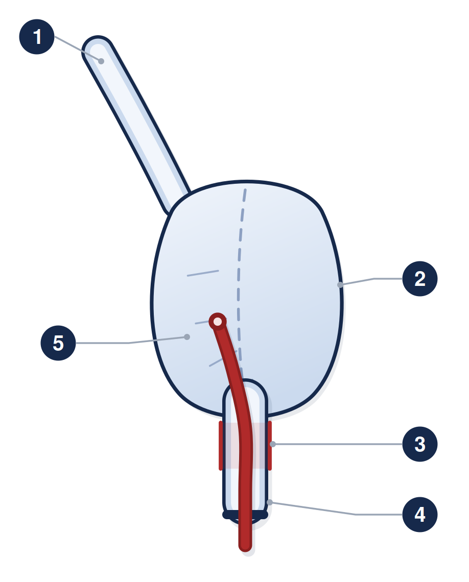
When the colon and rectum are removed, the end of the small bowel can be folded into a reservoir — a pouch — and joined to the anus, so that you open your bowels in the normal way. A short cuff of rectum is usually left behind at the join. A pouchoscopy uses a thin, flexible camera passed through the anus to look at the pouch, the cuff, and the small bowel just above the pouch.
It is done for two reasons. The first is to find the cause of symptoms — increased frequency, urgency, bleeding, pain or leakage — which may be inflammation of the pouch (pouchitis), inflammation of the cuff (cuffitis), a narrowing at the join, or Crohn’s-like disease of the pouch. The second is surveillance: taking small samples to look for early pre-cancerous change.
How often you need a surveillance pouchoscopy depends on your own situation — mainly whether any pre-cancerous change or cancer was found in the bowel that was removed, and whether you also have primary sclerosing cholangitis or long-standing pouch inflammation. For some people it is every year and for others every few years. Your team will write your own interval on this sheet. Cancer in the pouch is rare — fewer than 1 in every 100. What surveillance is actually looking for is the earlier, pre-cancerous change, which appears in about 2 in every 100 people over fifteen years and is treatable when it is caught. Both are much less likely if nothing pre-cancerous was found at your original operation.
1 the last part of the small bowel · 2 the pouch, made by folding the bowel back on itself · 3 the short cuff of rectum left at the join · 4 the anal canal · 5 the camera
Getting ready
You do not need the full bowel preparation used for a colonoscopy. An enema is usually enough, and the appointment letter will tell you when to use it — commonly about three hours before you leave home.
Have nothing to eat for six hours before, and clear fluids only up to two hours before.
Take your usual medicines unless you have been told otherwise. If you take insulin or tablets for diabetes, telephone the endoscopy unit first — the dose usually has to be adjusted around the fast.
Blood thinners: a diagnostic pouchoscopy with biopsies does not usually require them to be stopped, but tell the endoscopy unit what you take. If a narrowing is likely to need stretching, the plan may change — see sheet PE-PROC-16.
On the day
Most people have this test comfortably without any sedation. It is short — usually about ten minutes — and the pouch is close to the anus, so the camera does not have far to travel. You can choose gas and air, or sedation through a drip, if you would prefer. If you have sedation you will need a responsible adult to take you home and you must not drive for 24 hours; see sheet PE-PROC-17.
You will lie on your left side. Air or carbon dioxide is used to open the pouch so it can be seen properly, and small painless tissue samples are usually taken — from the small bowel above the pouch, from the pouch itself, and from the rectal cuff. The cuff samples matter, because that small piece of retained rectum is where pre-cancerous change is most likely to appear. Samples are taken above the sensitive part of the anal canal, which is why they do not hurt.
What are the risks?
Pouchoscopy is a short, low-risk examination and serious complications are very uncommon. There is no published figure for the complication rate of pouchoscopy specifically, so we quote the risks of flexible sigmoidoscopy, which is a comparable examination: bleeding after a biopsy and perforation of the bowel wall are both rare, in the region of 1 in 1,000 or fewer. Bloating and wind afterwards are expected rather than complications. If sedation is used, its own risks apply — see sheet PE-PROC-17.
If a narrowing at the join is stretched during the examination, that carries a higher risk and will be explained and consented separately.
Afterwards
You can eat and drink normally straight away unless you have been told otherwise, and go back to your usual activities the same day if you did not have sedation. Bloating, wind and a small amount of blood on wiping are common for a day or two.
The endoscopist will explain what was seen before you go home. Biopsy results take two to four weeks and are discussed at your next clinic appointment.
If pouchitis is found, it is usually treated with a two-week course of an antibiotic such as ciprofloxacin or metronidazole, and symptoms often settle within a couple of days. Pouchitis is common — around seven in every ten people with a pouch have at least one episode over the years. Of those who get it, more than half get it again, and about one in four of them develop a longer-lasting form that needs either regular antibiotics or a different kind of medicine. There are more treatment options for this now than there were a few years ago.
Get urgent help if
You have severe or worsening abdominal pain, or a hard, swollen abdomen.
You have a fever or shivering.
You are bleeding heavily from the back passage, or passing clots.
You feel faint or dizzy, or you collapse.
Take this sheet with you and tell the staff that you have had a pouchoscopy.
PE-PROC-06 · For appointments and questions, contact the endoscopy unit using the numbers it has given you. If you are seriously unwell, or it is outside working hours, go to the Emergency Department rather than waiting for a reply. This sheet supports the discussion with your doctor and the consent process; it does not replace them. Version 1.0 · review date October 2028.
This leaflet is general information and does not replace advice from your own doctor.